Download
1 / 13
130 likes | 709 Vues
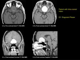
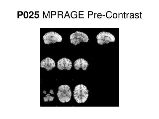
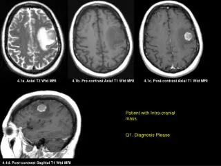
4.1a. Axial T2 Wtd MRI. 4.1b. Pre-contrast Axial T1 Wtd MRI. 4.1c. Post-contrast Axial T1 Wtd MRI. Patient with Intra-cranial mass. Q1. Diagnosis Please. 4.1d. Post-contrast Sagittal T1 Wtd MRI. 4.2a. Post-contrast Axial T1 Wtd MRI. 4.2b. Post-contrast Coronal T1 Wtd MRI.

E N D
4.1a. Axial T2 Wtd MRI 4.1b. Pre-contrast Axial T1 Wtd MRI 4.1c. Post-contrast Axial T1 Wtd MRI Patient with Intra-cranial mass. Q1. Diagnosis Please 4.1d. Post-contrast Sagittal T1 Wtd MRI
4.2a. Post-contrast Axial T1 Wtd MRI 4.2b. Post-contrast Coronal T1 Wtd MRI Patient with multiple Intra-cranial masses. Q2. Diagnosis Please
4.3a. Pre-contrast Axial T1 Wtd MRI 4.3b. Post-contrast Axial T1 Wtd MRI 4.3c. Post-contrast Coronal T1 Wtd MRI Abnormal MRI Q3. Diagnosis Please 4.3d. Post-contrast Sagittal T1 Wtd MRI
Abnormal MRI with dural masses Q4. Diagnosis Please 4.4. Post-contrast Coronal T1 Wtd MRI
Abnormal MRI Q5. Diagnosis Please 4.5. Post-contrast Axial T1 Wtd MRI
Fig. 4.1 Fig. 4.2 Fig. 4.3 Cases 4.1 through 4.5 share a common diagnosis. Name the common diagnosis. Fig. 4.4 Fig. 4.5
Fig. 4.1 Fig. 4.2 Fig. 4.3 Cases 4.1 through 4.5 share a common diagnosis. Name the common diagnosis. METASTASES TO THE BRAIN Fig. 4.4 Fig. 4.5
E 4.1a. Axial T2 Wtd MRI 4.1b. Pre-contrast Axial T1 Wtd MRI 4.1c. Post-contrast Axial T1 Wtd MRI 56-year old lady with a history of Breast carcinoma presented with 1-month history of increased difficulty in word finding. Findings: A well-defined enhancing tumor (arrow) is seen within the left frontal lobe with surrounding edema (E in figure A) Diagnosis: Solitary metastatic adenocarcinoma to the brain from breast primary. 4.1d. Post-contrast Sagittal T1 Wtd MRI
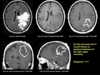
40-year old lady with a history of breast carcinoma diagnosed 6 years ago, presented with headache and ataxia. Findings: Shower of at least 30 metastatic enhancing lesions are seen closely packed together within both Cerebellar hemispheres (yellow arrows) and few lesions also seen within both posterior Fronto-parietal lobes (red arrows) Diagnosis: Multiple metastasis to the brain from breast primary
4.3a. Pre-contrast Axial T1 Wtd MRI 4.3b. Post-contrast Axial T1 Wtd MRI 4.3c. Post-contrast Coronal T1 Wtd MRI Findings: Linear enhancement of the subarachnoid space outlining the cerebellar sulci (arrows in B, C, D) and cortical sulci (small arrows in C,D). Note the pathology is not seen in non-contrast study (A). 28-year old male with melanoma presented with severe headaches, treated by chiropractor without relief, and also with blurred vision progressed to diplopia. Findings: Linear enhancement of the subarachnoid space outlining the cerebellar sulci (yellow arrows) and cortical sulci (red arrows). Note the pathology is not seen in non-contrast study (A). Diagnosis: Leptomeningeal/subarachnoid spread of melanoma metastasis, proven by cerebrospinal fluid cytology. 4.3d. Post-contrast Sagittal T1 Wtd MRI
69-year old male with prostate cancer diagnosed 7 years ago, presented with right sided hemiparesis. Findings: Multiple enhancing dural masses involving the left frontal dura (short arrow) and left temporal dura (long arrow) with calvarial metastases (red arrows). Diagnosis: Dural metastases from prostrate carcinoma 4.4. Post-contrast Coronal T1 Wtd MRI
50-year old male with renal cell carcinoma Diagnosis: Right temporal calvarial metastasis with a small epidural tumor (arrow head) and an intraventricular metastasis (arrows).
METASTASES TO THE BRAIN MRI is more sensitive than CT imaging to detect metastatic lesions and can detect a lesion as small as a dot (2mm). Common primary sites:Lung, Breast, Melanoma, Thyroid, Renal Cell Carcinoma • Patten of involvement: • Intraparenchymal (Figure 4.1) • Leptomeningial/Subarachnoid Spread (Figure 4.3) • Dural (Figure 4.4) • Epidural (Figure 4.5) • Intraventricular (Figure 4.5) Fig. 4.1 Fig. 4.3 • Hemorrhagic Metastases • Renal • Breast • Melanoma • Choriocracinoma Fig. 4.4 Fig. 4.5