Download
1 / 9
90 likes | 194 Vues
Learn about CNS lymphoma, rare brain tumors affecting corpus callosum, diagnostic imaging findings, and effective chemo-radiation therapy.

E N D
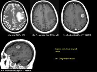
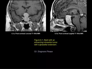
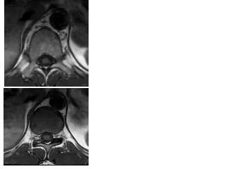
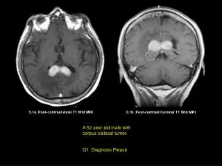
5.1a. Post-contrast Axial T1 Wtd MRI 5.1b. Post-contrast Coronal T1 Wtd MRI A 52 year-old male with corpus callosal tumor. Q1. Diagnosis Please
A 60 year-old male with known hemopoetic systemic disease. Q2. Diagnosis Please 5.2. Post-contrast Coronal T1 Wtd MRI
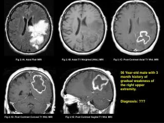
A 35 year-old female with multiple cranial nerve neuropathy. Q3. Diagnosis Please 5.3. Post-contrast Axial T1 Wtd MRI
5.1a. Post-contrast Axial T1 Wtd MRI 5.2. Post-contrast Coronal T1 Wtd MRI 5.3. Post-contrast Axial T1 Wtd MRI Figures 5.1 – 5.3 are produced by a hemopoetic tumor. Diagnosis: ?
5.1a. Post-contrast Axial T1 Wtd MRI 5.2. Post-contrast Coronal T1 Wtd MRI 5.3. Post-contrast Axial T1 Wtd MRI Figures 5.1 – 5.3 are produced by a hemopoetic tumor. Diagnosis: Lymphoma
5.1a. Post-contrast Axial T1 Wtd MRI 5.1b. Post-contrast Coronal T1 Wtd MRI Figure 5.1. Homogeneously enhancing tumor is seen involving the splenium of the corpus callosum (arrows) spreading across the midline. Diagnosis: Primary Lymphoma of the brain
Figure 5.2. Diagnosis: Secondary Lymphoma with calvarial involvement (green arrow) and associated epidural tumor (yellow arrows) / scalp tumor (red arrow) 5.2. Post-contrast Coronal T1 Wtd MRI
Figure 5.3. Linear enhancement of the cerebellar sulci (yellow arrows) and left temporal sulci (red arrow). Diagnosis: Secondary Lymphoma with subarachnoid tumor seeding (arrows). 5.3. Post-contrast Axial T1 Wtd MRI
Central Nervous System Lymphoma • Primary lymphoma of the brain is rare, accounts for less than 3.5% of primary brain tumors. • Non-Hodgkin’s Lymphoma, usually B-cell lymphoma. • Can be seen in patients with AIDS. • Tumor intensely enhances with contrast. Common sites of involvement include: • Basal ganglia/thalamus • Corpus Callosum • Periventricular white matter • Lymphoma responds well to chemo-radiation therapy. • Open or stereotactic biopsy is necessary to establish the diagnosis. • Complete surgical resection of the tumor is not necessary as they respond well to chemo-radiation therapy