Download
1 / 12
130 likes | 304 Vues
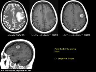
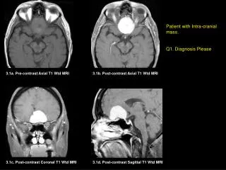
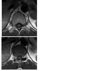
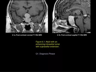
6.1a. Post-contrast coronal T1 Wtd MRI. 6.1b. Post-contrast sagittal T1 Wtd MRI. Figure 6.1: Adult with an enhancing intrasellar tumor with suprasellar extension. Q1. Diagnosis Please. 6.2. Post-contrast sagittal T1 Wtd MRI. Figure 6.2: A child with cystic suprasellar tumor. .

E N D
6.1a. Post-contrast coronal T1 Wtd MRI 6.1b. Post-contrast sagittal T1 Wtd MRI Figure 6.1: Adult with an enhancing intrasellar tumor with suprasellar extension. Q1. Diagnosis Please
6.2. Post-contrast sagittal T1 Wtd MRI Figure 6.2: A child with cystic suprasellar tumor. Q2. Diagnosis Please
6.3a. Post-contrast sagittal T1 Wtd MRI 6.3b. Post-contrast axial T1 Wtd MRI Figure 6.3: A child with nausea, vomiting and ataxia. Q3. Diagnosis Please
6.4a. Post-contrast axial T1 Wtd MRI 6.4b. Post-contrast axial T1 Wtd MRI 6.4c. Pre-contrast sagittal T1 Wtd MRI Figure 6.4: A child with nausea, vomiting and ataxia. Q4. Diagnosis Please
6.5a. Pre-contrast axial T1 Wtd MRI 6.5b. Post-contrast axial T1 Wtd MRI 6.5c. Post-contrast coronal T1 Wtd MRI Figure 6.5: A child with nausea, vomiting and ataxia. Q5. Diagnosis Please
6.6a. Pre-contrast axial T1 Wtd MRI 6.6b. Post-contrast axial T1 Wtd MRI Figure 6.6: An adult with a cranial nerve deficit produced by a tumor. Name the cranial nerve deficit _____________ Name the tumor ____________ Q6. Diagnosis Please
6.1a. Post-contrast coronal T1 Wtd MRI 6.1b. Post-contrast sagittal T1 Wtd MRI Figure 6.1 Findings: A homogeneously enhancing tumor is seen within the sella (yellow arrow in figures A, B) with suprasellar extension (red arrow in figures A, B) producing optic chiasm compression. Diagnosis: • PITUITARY ADENOMA • Non-hormone secreting pituitary macro adenomas greater than 1cm in size, grow silently until they produce optic chiasm compression giving rise to visual field defects. • Hormone secreting tumors such as eosinophilic adenoma giving rise to gigantism/acromegaly and ACTH producing tumor resulting in Cushing’s Syndrome are usually smaller in size measuring less than 1cm in size (microadenoma). Microadenomas tend to be small in size and are recognized early due to the hormone secreting features.
6.2. Post-contrast sagittal T1 Wtd MRI Figure 6.2 Findings: A large cystic suprasellar tumor (yellow arrow) with a rim of peripheral enhancement (red arrow). Diagnosis: • CRANIOPHARYNGIOMA • Common suprasellar tumor in a child • Tumors can be cystic/solid • Common calcified tumor, calcification within the tumor can be seen as high as 90%
6.3a. Post-contrast sagittal T1 Wtd MRI 6.3b. Post-contrast axial T1 Wtd MRI Figure 6.3 Findings: An enhancing intra IV ventricular tumor (yellow arrow in figures A, B) is seen. Red arrows point to expanded IV ventricle. Diagnosis: • EPENDYMOMA OF THE IV VENTRICLE • Common pediatric brain tumor • Enhancing tumors. Calcification can be seen in 50% of tumors.
T 6.4a. Pre-contrast axial T1 Wtd MRI 6.4b. Post-contrast axial T1 Wtd MRI 6.4c. Pre-contrast sagittal T1 Wtd MRI Figure 6.4 Findings: An enhancing tumor (yellow arrow in B) seen posterior to the IV ventricle. Pre-contrast sagittal T1-weighted MR image (figure c) shows tumor (T) and its location posterior inferior to the IV ventricle (yellow arrow). Diagnosis: • MEDULLOBLASTOMA • Common pediatric brain tumor • Common location is posterior to the IV ventricle, involving the vermis. • Tumor enhances with contrast.
6.5a. Pre-contrast axial T1 Wtd MRI 6.5b. Post-contrast axial T1 Wtd MRI 6.5c. Post-contrast coronal T1 Wtd MRI Figure 6.5 Findings: Left cerebellar tumor with cystic (yellow arrow in A,B,C) and a solid enhancing tumor nodule (red arrow in B,C). Diagnosis: • JUVENILE PILOCYTIC. ASTROCYTOMA • Common pediatric brain tumor • Common locations include cerebellum and cerebral hemispheres. • Tumors demonstrate solid and cystic components
6.6a. Pre-contrast axial T1 Wtd MRI 6.6b. Post-contrast axial T1 Wtd MRI Figure 6.6 Findings: An enhancing tumor within the left cerebello-pontine angel (red arrow) with extension into the internal auditory canal (yellow arrow) Diagnosis: • Vestibular (8th nerve) Schwannoma • Tumor recognized by its location. • Tumor intensely enhances with contrast.