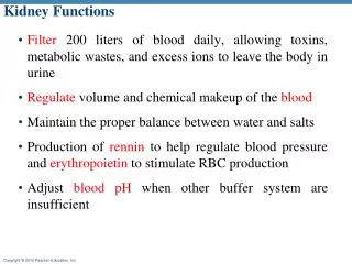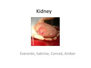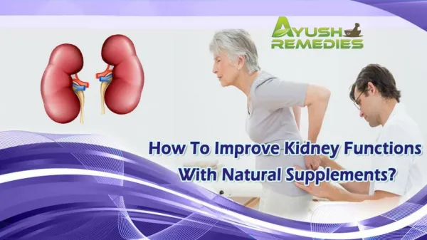
Kidney Functions
Learn about the intricate functions and anatomy of the kidneys, including how they filter blood, regulate blood volume and chemical makeup, and produce essential hormones. Explore the renal system's organs and layers of tissue supporting the kidneys.

Kidney Functions
E N D
Presentation Transcript
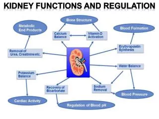
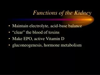
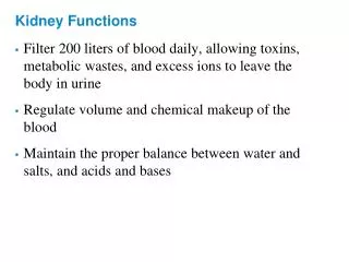
Kidney Functions • Filter 200 liters of blood daily, allowing toxins, metabolic wastes, and excess ions to leave the body in urine • Regulate volume and chemical makeup of the blood • Maintain the proper balance between water and salts, and acids and bases
Other Renal Functions • Gluconeogenesis during prolonged fasting • Production of rennin to help regulate blood pressure and erythropoietin to stimulate RBC production • Activation of vitamin D
Other Urinary System Organs • Urinary bladder – provides a temporary storage reservoir for urine • Paired ureters – transport urine from the kidneys to the bladder • Urethra – transports urine from the bladder out of the body
Kidney Location and External Anatomy • The kidneys lie in a retroperitoneal position in the superior lumbar region • The right kidney is lower than the left because it is crowded by the liver • The lateral surface is convex; the medial surface is concave • The renal hilus leads to the renal sinus • Ureters, renal blood vessels, lymphatics, and nerves enter and exit at the hilus
Layers of Tissue Supporting the Kidney • Renal capsule – fibrous capsule that prevents kidney infection • Adipose capsule – fatty mass that cushions the kidney and helps attach it to the body wall • Renal fascia – outer layer of dense fibrous connective tissue that anchors the kidney
Internal Anatomy (Frontal Section) • Cortex – the light colored, granular superficial region • Medulla – exhibits cone-shaped medullary (renal) pyramids separated by columns • The medullary pyramid and its surrounding capsule constitute a lobe • Renal pelvis – flat funnel shaped tube lateral to the hilus within the renal sinus
Internal Anatomy • Major calyces – large branches of the renal pelvis • Collect urine draining from papillae • Empty urine into the pelvis • Urine flows through the pelvis and ureters to the bladder
Blood and Nerve Supply • Approximately one-fourth (1200 ml) of systemic cardiac output flows through the kidneys each minute • Arterial flow into and venous flow out of the kidneys follow similar paths • The nerve supply is via the renal plexus
Renal Vascular Pathway Figure 25.3c
The Nephron • Nephrons are the structural and functional units that form urine, consisting of: • Glomerulus – a tuft of capillaries associated with a renal tubule • Glomerular (Bowman’s) capsule – blind, cup-shaped end of a renal tubule that completely surrounds the glomerulus
The Nephron • Renal corpuscle – the glomerulus and its Bowman’s capsule • Glomerular endothelium – fenestrated epithelium that allows solute-rich, virtually protein-free filtrate to pass from the blood into the glomerular capsule
Anatomy of the Glomerular Capsule • The external parietal layer is a structural layer • The visceral layer consists of modified, branching epithelial podocytes • Extensions of the octopus-like podocytes terminate in foot processes • Filtration slits – openings between the foot processes that allow filtrate to pass into the capsular space
Renal Tubule • Proximal convoluted tubule (PCT) – composed of cuboidal cells with numerous microvilli and mitochondria • Reabsorbs water and solutes from filtrate and secretes substances into it
Renal Tubule • Loop of Henle – a hairpin-shaped loop of the renal tubule • Proximal part is similar to the proximal convoluted tubule • Proximal part is followed by the thin segment (simple squamous cells) and the thick segment (cuboidal to columnar cells) • Distal convoluted tubule (DCT) – cuboidal cells without microvilli that function more in secretion than reabsorption
Connecting Tubules • The distal portion of the distal convoluted tubule nearer to the collecting ducts
Connecting Tubules • Two important cell types are found here • Intercalated cells • Cuboidal cells with microvilli • Function in maintaining the acid-base balance of the body • Principal cells • Cuboidal cells without microvilli • Help maintain the body’s water and salt balance
Nephrons • Cortical nephrons – 85% of nephrons; located in the cortex • Juxtamedullary nephrons: • Are located at the cortex-medulla junction • Have loops of Henle that deeply invade the medulla • Have extensive thin segments • Are involved in the production of concentrated urine
Capillary Beds of the Nephron • Every nephron has two capillary beds • Glomerulus • Peritubular capillaries • Each glomerulus is: • Fed by an afferent arteriole • Drained by an efferent arteriole
Capillary Beds of the Nephron • Blood pressure in the glomerulus is high because: • Arterioles are high-resistance vessels • Afferent arterioles have larger diameters than efferent arterioles • Fluids and solutes are forced out of the blood throughout the entire length of the glomerulus
Capillary Beds • Peritubular beds are low-pressure, porous capillaries adapted for absorption that: • Arise from efferent arterioles • Cling to adjacent renal tubules • Empty into the renal venous system • Vasa recta – long, straight efferent arterioles of juxtamedullary nephrons
Vascular Resistance in Microcirculation • Afferent and efferent arterioles offer high resistance to blood flow • Blood pressure declines from 95mm Hg in renal arteries to 8 mm Hg in renal veins
Vascular Resistance in Microcirculation • Resistance in afferent arterioles: • Protects glomeruli from fluctuations in systemic blood pressure • Resistance in efferent arterioles: • Reinforces high glomerular pressure • Reduces hydrostatic pressure in peritubular capillaries
Juxtaglomerular Apparatus (JGA) • Where the distal tubule lies against the afferent (sometimes efferent) arteriole • Arteriole walls have juxtaglomerular (JG) cells • Enlarged, smooth muscle cells • Have secretory granules containing renin • Act as mechanoreceptors
Juxtaglomerular Apparatus (JGA) • Macula densa • Tall, closely packed distal tubule cells • Lie adjacent to JG cells • Function as chemoreceptors or osmoreceptors • Mesanglial cells: • Have phagocytic and contractile properties • Influence capillary filtration
Filtration Membrane • Filter that lies between the blood and the interior of the glomerular capsule • It is composed of three layers • Fenestrated endothelium of the glomerular capillaries • Visceral membrane of the glomerular capsule (podocytes) • Basement membrane composed of fused basal laminae of the other layers
Mechanisms of Urine Formation • The kidneys filter the body’s entire plasma volume 60 times each day • The filtrate: • Contains all plasma components except protein • Loses water, nutrients, and essential ions to become urine • The urine contains metabolic wastes and unneeded substances
Mechanisms of Urine Formation • Urine formation and adjustment of blood composition involves three major processes • Glomerular filtration • Tubular reabsorption • Secretion Figure 25.8
Glomerular Filtration • Principles of fluid dynamics that account for tissue fluid in all capillary beds apply to the glomerulus as well • The glomerulus is more efficient than other capillary beds because: • Its filtration membrane is more permeable • Glomerular blood pressure is higher • It has a higher net filtration pressure • Plasma proteins are not filtered and are used to maintain pressure of the blood
Net Filtration Pressure (NFP) • The pressure responsible for filtrate formation • NFP equals the glomerular hydrostatic pressure (HPg) minus the oncotic pressure of glomerular blood (OPg) combined with the capsular hydrostatic pressure (HPc) NFP = HPg – (OPg +HPc)
Glomerular Filtration Rate (GFR) • The total amount of filtrate formed per minute by the kidneys • Factors governing filtration rate at the capillary bed are: • Total surface area available for filtration • Filtration membrane permeability • Net filtration pressure
Glomerular Filtration Rate (GFR) • GFR is directly proportional to the NFP • Changes in GFR normally result from changes in glomerular blood pressure
Regulation of Glomerular Filtration • If the GFR is too high: • Needed substances cannot be reabsorbed quickly enough and are lost in the urine • If the GFR is too low: • Everything is reabsorbed, including wastes that are normally disposed of
Regulation of Glomerular Filtration • Three mechanisms control the GFR • Renal autoregulation (intrinsic system) • Neural controls • Hormonal mechanism (the renin-angiotensin system)
Intrinsic Controls • Under normal conditions, renal autoregulation maintains a nearly constant glomerular filtration rate • Autoregulation entails two types of control • Myogenic – responds to changes in pressure in the renal blood vessels • Flow-dependent tubuloglomerular feedback – senses changes in the juxtaglomerular apparatus
Extrinsic Controls • When the sympathetic nervous system is at rest: • Renal blood vessels are maximally dilated • Autoregulation mechanisms prevail
Extrinsic Controls • Under stress: • Norepinephrine is released by the sympathetic nervous system • Epinephrine is released by the adrenal medulla • Afferent arterioles constrict and filtration is inhibited • The sympathetic nervous system also stimulates the renin-angiotensin mechanism
Renin-Angiotensin Mechanism • Is triggered when the JG cells release renin • Renin acts on angiotensinogen to release angiotensin I • Angiotensin I is converted to angiotensin II • Angiotensin II: • Causes mean arterial pressure to rise • Stimulates the adrenal cortex to release aldosterone • As a result, both systemic and glomerular hydrostatic pressure rise
Renin Release • Renin release is triggered by: • Reduced stretch of the granular JG cells • Stimulation of the JG cells by activated macula densa cells • Direct stimulation of the JG cells via 1-adrenergic receptors by renal nerves • Angiotensin II