Kidney Functions
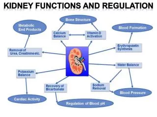
Kidney Functions. Filter 200 liters of blood daily, allowing toxins, metabolic wastes, and excess ions to leave the body in urine Regulate volume and chemical makeup of the blood Maintain the proper balance between water and salts, and acids and bases. Other Renal Functions.

Kidney Functions
E N D
Presentation Transcript
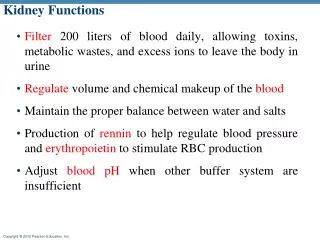
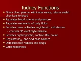
Kidney Functions • Filter 200 liters of blood daily, allowing toxins, metabolic wastes, and excess ions to leave the body in urine • Regulate volume and chemical makeup of the blood • Maintain the proper balance between water and salts, and acids and bases
Other Renal Functions • Gluconeogenesis during prolonged fasting • Production of rennin to help regulate blood pressure and erythropoietin to stimulate RBC production • Activation of vitamin D
Other Urinary System Organs • Urinary bladder – provides a temporary storage reservoir for urine • Paired ureters – transport urine from the kidneys to the bladder • Urethra – transports urine from the bladder out of the body
Urinary System Organs Figure 25.1a
Kidney Location and External Anatomy • The kidneys lie in the superior lumbar region, deep within the body. • The right kidney is lower than the left because it is crowded by the liver • The lateral surface is convex; the medial surface is concave • The renal hilus leads to the renal sinus • Ureters, renal blood vessels, lymphatics, and nerves enter and exit at the hilus
Kidney Location and External Anatomy Figure 25.2a
Internal Anatomy Figure 25.3b
The Nephron • Nephrons are the structural and functional units that form urine, consisting of: • Glomerulus – a tuft of capillaries associated with a renal tubule • Glomerular (Bowman’s) capsule – blind, cup-shaped end of a renal tubule that completely surrounds the glomerulus
The Nephron • Renal corpuscle – the glomerulus and its Bowman’s capsule • Glomerular endothelium – fenestrated epithelium that allows solute-rich, virtually protein-free filtrate to pass from the blood into the glomerular capsule
Posterior vena cava Renal artery and vein Kidney Aorta Ureter Urinary bladder Urethra (a) Excretory organs and major associated blood vessels Kidneys • Concept 44.4: Nephrons and associated blood vessels are the functional unit of the kidney
Overview: A Balancing Act • Physiological systems of animals operate in a fluid environment • Relative concentrations of water and solutes must be maintained within fairly narrow limits • Osmoregulation regulates solute concentrations and balances the gain and loss of water
Concept 44.1: Osmoregulation balances the uptake and loss of water and solutes • Osmoregulation is based largely on controlled movement of solutes between internal fluids and the external environment Recall: Movement of water is osmosis Movement of solutes is diffusion To get water to flow BACK to the right, you could add solutes to the right side, increasing the concentration of concentration of solutes, thus decreasing the concentration of water. Water would then move from left (higher water concentration) to the right (lower water concentration) Your kidneys do the same thing.
Concept 44.2: An animal’s nitrogenous wastes reflect its phylogeny and habitat • The type and quantity of an animal’s waste products may greatly affect its water balance • Among the most important wastes that are removed by the kidneys are nitrogenous breakdown products of proteins and nucleic acids • Some animals convert toxic ammonia (NH3) to less toxic compounds prior to excretion
Fig. 44-9a Most aquatic animals, including most bony fishes Many reptiles (including birds), insects, land snails Mammals, most amphibians, sharks, some bony fishes Ammonia Urea Uric acid
Urea • The liver of mammals and most adult amphibians converts ammonia to urea • The circulatory system carries urea to the kidneys, where it is excreted • Conversion of ammonia to urea is energetically expensive; excretion of urea requires less water than ammonia
Excretory Processes • The human excretory system produces urine by refining a filtrate derived from body fluids • Key functions of most excretory systems: • Filtration: pressure-filtering of body fluids • Reabsorption: reclaiming valuable solutes: i.e., putting it back into the blood • Secretion: adding toxins and other solutes from the body fluids to the filtrate • Excretion: removing the filtrate from the system
Fig. 44-10 Filtration Capillary Filtrate Excretory tubule Reabsorption Secretion Urine Excretion
Structure of the Mammalian Excretory System • The mammalian excretory system centers on paired kidneys, which are also the principal site of water balance and salt regulation • Each kidney is supplied with blood by a renal artery and drained by a renal vein • Urine exits each kidney through a duct called the ureter • Both ureters drain into a common urinary bladder, and urine is expelled through a urethra
The mammalian kidney has two distinct regions: an outer renal cortex and an inner renal medulla
Fig. 44-14b Renal medulla Renal cortex Renal pelvis Ureter Section of kidney from a rat (b) Kidney structure 4 mm
Fig. 44-14cd Afferent arteriole from renal artery Glomerulus Juxtamedullary nephron Cortical nephron Bowman’s capsule 10 µm SEM Proximal tubule Peritubular capillaries Renal cortex Efferent arteriole from glomerulus Collecting duct Distal tubule Branch of renal vein Renal medulla Collecting duct Descending limb To renal pelvis Loop of Henle Ascending limb Vasa recta (c) Nephron types (d) Filtrate and blood flow
The nephron, the functional unit of the vertebrate kidney, consists of a single long tubule and a ball of capillaries called the glomerulus • Bowman’s capsule surrounds and receives filtrate from the glomerulus
Fig. 44-14c Juxtamedullary nephron Cortical nephron Renal cortex Collecting duct Renal medulla To renal pelvis (c) Nephron types
Fig. 44-14d Glomerulus Afferent arteriole from renal artery Bowman’s capsule 10 µm SEM Proximal tubule Peritubular capillaries Efferent arteriole from glomerulus Distal tubule Branch of renal vein Collecting duct Descending limb Loop of Henle Ascending limb Vasa recta (d) Filtrate and blood flow
Filtration of the Blood • Filtration occurs as blood pressure forces fluid from the blood in the glomerulus into the lumen of Bowman’s capsule • Filtration of small molecules is nonselective • The filtrate contains salts, glucose, amino acids, vitamins, nitrogenous wastes, and other small molecules
Pathway of the Filtrate • From Bowman’s capsule, the filtrate passes through three regions of the nephron: the proximal tubule, the loop of Henle, and the distal tubule • Fluid from several nephrons flows into a collecting duct, all of which lead to the renal pelvis,which is drained by the ureter • Cortical nephrons are confined to the renal cortex, while juxtamedullary nephrons have loops of Henle that descend into the renal medulla
Blood Vessels Associated with the Nephrons • Each nephron is supplied with blood by an afferent arteriole, a branch of the renal artery that divides into the capillaries • The capillaries converge as they leave the glomerulus, forming an efferent arteriole • The vessels divide again, forming the peritubular capillaries, which surround the proximal and distal tubules
Concept 44.4: The nephron is organized for stepwise processing of blood filtrate • The mammalian kidney conserves water by producing urine that is much more concentrated than body fluids
From Blood Filtrate to Urine: A Closer Look Proximal Tubule • Reabsorption of ions, water, and nutrients takes place in the proximal tubule • Molecules are transported actively and passively from the filtrate into the interstitial fluid and then capillaries • Some toxic materials are secreted into the filtrate • The filtrate volume decreases Descending Limb of the Loop of Henle Reabsorption of water continues through channels formed by aquaporin proteins Movement is driven by the high osmolarity of the interstitial fluid, which is hyperosmotic to the filtrate The filtrate becomes increasingly concentrated
Ascending Limb of the Loop of Henle • In the ascending limb of the loop of Henle, salt but not water is able to diffuse from the tubule into the interstitial fluid • The filtrate becomes increasingly dilute • Distal Tubule • The distal tubule regulates the K+ and NaCl concentrations of body fluids • The controlled movement of ions contributes to pH regulation • Collecting Duct • The collecting duct carries filtrate through the medulla to the renal pelvis • Water is lost as well as some salt and urea, and the filtrate becomes more concentrated
Click on the image below to see a tutorial on macroscopic anatomy of the kidney.
Click on the image below to watch a tutorial on the physiology of the nephron
Antidiuretic Hormone • The osmolarity of the urine is regulated by nervous and hormonal control of water and salt reabsorption in the kidneys • Antidiuretic hormone (ADH) increases water reabsorption in the distal tubules and collecting ducts of the kidney • An increase in osmolarity triggers the release of ADH, which helps to conserve water
The Renin-Angiotensin-Aldosterone System • The renin-angiotensin-aldosterone system (RAAS) is part of a complex feedback circuit that functions in homeostasis • A drop in blood pressure near the glomerulus causes the juxtaglomerular apparatus (JGA) to release the enzyme renin • Renin triggers the formation of the peptide angiotensin II
Angiotensin II • Raises blood pressure and decreases blood flow to the kidneys • Stimulates the release of the hormone aldosterone, which increases blood volume and pressure
Click on the image below to watch a tutorial on the RAAS system:
Juxtaglomerular Apparatus (JGA) • Has mechanoreceptors that can detect increased blood pressure • Has cells that can release renin