Download
1 / 8
80 likes | 277 Vues
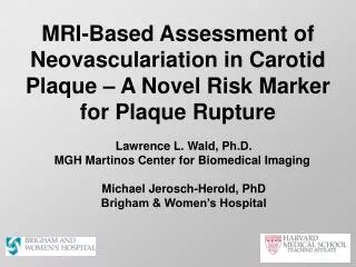
MRI-Based Assessment of Neovasculariation in Carotid Plaque – A Novel Risk Marker for Plaque Rupture. Michael Jerosch-Herold, PhD Associate Professor of Radiology Director of Physics for the Cardiac Imaging Program Brigham & Women’s Hospital.

E N D
MRI-Based Assessment of Neovasculariation in Carotid Plaque – A Novel Risk Marker for Plaque Rupture Michael Jerosch-Herold, PhD Associate Professor of Radiology Director of Physics for the Cardiac Imaging Program Brigham & Women’s Hospital
New 3.0 T Bilateral 8-Channel Carotid Array and Head-Holder for Reynold Carotid Study Head Holder MGH Design Wiggins/Wald Reference: Hinton-Yates et al., TMRI 2007 8- Coils = pair of four 4.8 cm loops Coil mounted on a frame with head-holder consists of two curved paddles
MRI Protocol for Dynamic Contrast Enhancement • T1-weighted 3D fast gradient echo • TR/TE/flip angle: 4.3/2.3 ms/ 20° • 3 mm slices • In-plane resolution: 0.7 x 0.7 mm • Time per dynamic view: 14 s • With new 8-element neck phased array the time per dynamic view is reduced to 10 s.
Post-Processing Contrast-Enhancement in Carotid Wall
Ktrans Parametric Map 24 20 16 Ktrans*1e3 [1/min] 12 8 4 Kinetic Modeling(Kety-Schmidt Model) Measured in vessel lumen Venous Outflow Arterial Input vb Tissue Extraction (Ktrans) • Model Parameters Determined by Optimization at Pixel Level • vb vascular volume • Ktrans First order blood-to-tissue transfer rate constant
Ktrans Parametric Map 24 20 16 Ktrans*1e3 [1/s] 12 8 4 Examp T2 TSE: carotid plaque
Example of Test-Scan in Patient with 8-Channel Phased Array Coil