The Brain Module 5
The Brain Module 5. The Brain. The Brain The Tools of Discovery Older Brain Structures The Cerebral Cortex Our Divided Brain Left Brain-Right Brain. The Brain. Techniques to Study the Brain.

The Brain Module 5
E N D
Presentation Transcript
The Brain The Brain • The Tools of Discovery • Older Brain Structures • The Cerebral Cortex • Our Divided Brain • Left Brain-Right Brain
The Brain Techniques to Study the Brain Brain lesion experimentally destroys brain tissue to study animal behaviors after such destruction. Hubel (1990)
Clinical Observation Neurological and psychiatric diseases are now catalogued. Tom Landers/ Boston Globe
Electroencephalogram (EEG) An amplified recording of the electrical waves sweeping across the brain’s surface, measured by electrodes placed on the scalp. AJ Photo/ Photo Researchers, Inc.
PET Scan PET (positron emission tomography) Scan a visual display of brain activity that detects a radioactive form of glucose while the brain performs a given task. Courtesy of National Brookhaven National Laboratories
MRI & fMRI Scan ventricular enlargement in a schizophrenic patient. MRI (magnetic resonance imaging) uses magnetic fields and radio waves that reveal structure. fMRI (functional MRI) reveals brain’s functioning by making a sort of “movie” of changes in the activity of the brainand structure. Both photos from Daniel Weinberger, M.D., CBDB, NIMH James Salzano/ Salzano Photo Lucy Reading/ Lucy Illustrations brain regions when a participant lies.
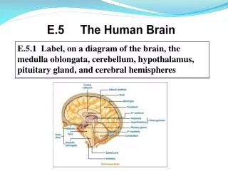
Older Brain Structures Brainstemthe oldest part of the brain, beginning where the spinal cord swells and enters the skull. Responsible for automatic survival functions.
Brain Stem Medulla [muh-DUL-uh] base of the brainstem, controls heartbeat and breathing. Reticular Formation – a nerve network inside the brainstem that plays an important role in controlling arousal.
Brain Stem Thalamus [THAL-uh-muss] the brain’s sensory switchboard, located on top of the brainstem. It directs messages to the sensory areas in the cortex and transmits replies to the cerebellum and medulla.
Cerebellum The “little brain” attached to the rear of the brainstem. It helps coordinate voluntary movements and balance along with some nonverbal learning and memory.
The Limbic System Limbic Systema doughnut-shaped system of neural structures at the border of the brainstem and cerebrum, associated with emotions such as fear, aggression and drives for food and sex. It includes the hippocampus, amygdala, and hypothalamus.
Amygdala Amygdala [ah-MIG-dah-la] two almond-shaped neural clusters linked to emotion of fear and anger.
Hypothalamus Hypothalamus lies below (hypo) the thalamus; directs several maintenance activities like eating, drinking body temperature, and emotions. Helps govern the endocrine system via the pituitary gland.
Reward Center (nucleus accumbens) Rats cross an electrified grid for self-stimulation, when electrodes are placed in the reward center (hypothalamus) . When the limbic system is manipulated rat will navigate fields or climb up a tree. Sanjiv Talwar, SUNY Downstate
The Cerebral Cortex The intricate fabric of interconnected neural cells that covers the cerebral hemispheres. The body’s ultimate control and information processing center.
Structure of the Cortex frontal lobes (forehead) – planning, organization, impulse control parietal lobes (top to rear head) – touch, taste, temperature occipital lobes (back head) – vision temporal lobes (side of head) – auditory and higher visual processing.
Functions of the Cortex Motor Cortex area at the rear of the frontal lobes controls voluntary movements. Sensory Cortex (parietal cortex) receives information from skin surface and sense organs.
Visual Function Functional MRI scan shows the visual cortex activates as the subject looks at faces. Courtesy of V.P. Clark, K. Keill, J. Ma. Maisog, S. Courtney, L.G. Ungerleider, and J.V. Haxby, National Institute of Mental Health
Auditory Function Functional MRI scan shows the auditory cortex is active in patients who hallucinate.
Association Areas More intelligent animals have increased “uncommitted” or association areas of the cortex.
Spatial neglect - damage to the association areas of the right hemisphere resulting in an inability to recognize objects or body parts in the left visual field.
Language Aphasiais an impairment of language, usually caused by left hemisphere damage either to Broca’s area (impaired speaking) or to Wernicke’s area (impaired understanding).
LO 2.13 Left side and right side of brain Language is primarily a left hemisphere activity for most individuals Menu
Specialization & Integration Brain activity when hearing, seeing, and speaking words
The Brain’s Plasticity Brain is sculpted by our genes but also by our experiences. Plasticityrefers to the brain’s ability to modify itself after some type of injury or illness.
Our Divided Brain – two hemispheres Left hemisphere - Sequential processing for analytic reasoning and language, reading, writing, calculations, comprehension skills, and thus termed as the dominant brain in the 1960s. Right hemisphere – Simultaneous processing, nonverbal, visual-spatial, melody, pitch, emotional content of language
Splitting the Brain A procedure in which the two hemispheres of the brain are isolated by cutting the connecting fibers (mainly those of the corpus callosum) between them. Corpus Callosum Courtesy of Terence Williams, University of Iowa Martin M. Rother
Split Brain Patients With the corpus callosum severed, objects (apple) presented in the right visual field can be named. Objects (pencil) in the left visual field cannot.
LO 2.13 Left side and right side of brain Split-brain subjects stared at a dot and viewed a composite of two faces (A). When asked what they saw, subjects chose the child—the image sent to the verbal left hemisphere (B). But when subjects pointed to the face with the left hand, they chose the woman with glasses—whose image was received by the right hemisphere (C) (Levy et al., 1983). Menu
Try This! Try drawing two shapes with both of you hands simultaneously. BBC
Non-Split Brains People with intact brains also show left-right hemispheric differences in mental abilities. A number of brain scan studies have shown normal individuals engage their right brain when they engage in a perceptual task, and left brain when carrying out a linguistic task.
Brain Organization & Handedness Is handedness inherited? Yes. Archival and historic studies to modern medical studies point that right hand is preferred. This suggests, genes and/or prenatal factors influence handedness.
Is it All Right to be Left Handed? Being a left hander is difficult in a right-handed world.
Is it All Right to be Left Handed? The percentage of left-handers decreases sharply in samples of older people (Coren, 1993).