Download
1 / 17
170 likes | 285 Vues
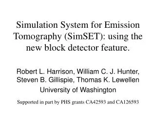
Simulation System for Emission Tomography (SimSET): using the new block detector feature. Robert L. Harrison, William C. J. Hunter, Steven B. Gillispie, Thomas K. Lewellen University of Washington Supported in part by PHS grants CA42593 and CA126593. What is SimSET?.

E N D
Simulation System for Emission Tomography (SimSET): using the new block detector feature. Robert L. Harrison, William C. J. Hunter, Steven B. Gillispie, Thomas K. Lewellen University of Washington Supported in part by PHS grants CA42593 and CA126593
What is SimSET? • A modular photon-tracking simulation. • PET and SPECT. • Public domain. • Over 400 registered users. • Used in over 250 papers/dissertations/theses.
What is SimSET used for? • Optimizing patient studies. • Assessing and improving quantitation. • Prototyping tomographs.
Block detector model • More faithful model of typical PET scanners. • More realistic simulated results, both quantitatively and qualitatively.
Simulating a block detector tomograph • General Electric DSTE PET scanner. • Steps in defining a simulation: • Phantom. • Collimator. • Detector(s). • Desired output.
Phantom • NEMA NU2-2001 scatter phantom. • The dimensions are given in the run parameters file. • Specify activity and attenuation for each voxel.
DSTE collimators • 3D mode. • SimSET models PET collimators as cylindrical annuli. • Endplates are the main feature. • Also included patient tunnel and front face of detector housing.
Defining the detector array • Define blocks. • Arrange blocks into detector rings. • Stack rings.
DSTE blocks • Dimensions. • Material. • Active (e.g. scintillating) or not?
DSTE rings • Position blocks. • Different types of blocks were used to represent: • Block housing and support. • Interblock shielding. • etc.
DSTE tomograph • Stack rings axially. • Different types of rings were included to represent material outside the detector rings. • Specify energy resolution.
Defining the output • A bin for every crystal pair: • 24 axial slices of crystals. • 560 crystals per ring. • SimSET can also bin by detected energy, and by true/scatter/random state.
Experimental method • Using measured and simulated data: • Computed scatter fraction. • Created images of single photon distribution. • Performed simulations using: • DSTE block detector array presented here. • More detailed block detector DSTE simulation. • A cylindrical representation of the DSTE using SimSET’s old cylindrical detector model.
Singles distribution for NEMANU2-2001 phantom Experimental vs. simulations: detected single events histogrammed by axial ring and crystal within the ring.
Summary • SimSET’s new block detector module significantly improves the accuracy of tomograph modeling. • Accurate modeling required for best results.
For further information • Website: • http://depts.washington.edu/simset/html/simset_main.html • Contact: • simset@u.washington.edu