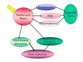
NUCLEIC ACID
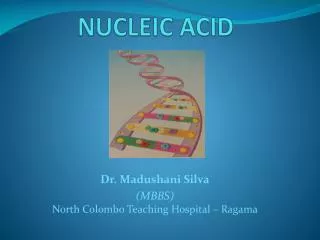
NUCLEIC ACID. Dr. Madushani Silva (MBBS) North Colombo Teaching Hospital – Ragama. Nucleic Acids ( DNA / RNA). Why are we studying Nucleic acids ??? . Passing characteristics of animal from generation to generation. . Store all the information needed to make us what we are .

NUCLEIC ACID
E N D
Presentation Transcript
NUCLEIC ACID Dr. Madushani Silva (MBBS)North Colombo Teaching Hospital – Ragama
Nucleic Acids ( DNA / RNA) Why are we studying Nucleic acids ??? Passing characteristics of animal from generation to generation. Store all the information needed to make us what we are Because they play a vital role in. Mistakes in them cause genetic diseases.
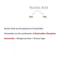
Deoxyribonucleic acid. (DNA) Nucleic acids Ribonucleic acid (RNA) Nucleoside Sugar + N containing base Nucleotide Nucleoside + Phosphate Nucleic acids = Polymers of Nucleotides Polynucleotides
Components of Nucleic acids. 5 HOCH2 OH O 4 1 1). 5 carbon sugar. 2 3 OH OH
2). The N containing bases of Nucleic acids Pyrimidines (single ring) Purines (double rings) Cytosine Thymine Uracil (only in RNA) Adinine Guanine
The N containing bases of Nucleic acids
5 HOCH2 OH O Add a purine / pyrimidine base 4 1 2 3 OH OH Ribose 5 HOCH2 BASE O 4 1 2 3 OH OH Nucleoside
Add a phosphate to a nucleoside Nucleotide ‒ O I O = P – O I O 5 CH2 BASE O 4 1 ‒ 2 3 OH OH A Nucleotide
Nucleotides link together by phosphodiester links ‒ O I O = P – O I O 5 CH2 BASE O 4 1 ‒ 2 3 OH O I O = P – O I O 5 Phospho- diester link (5’ 3’) CH2 BASE O 4 1 ‒ 2 3 OH OH
A closer look at the polynucleotide chain Note * 3’ end and 5’ end of chain * Sugar – phosphate backbone * Bases stick out
The simple ways in which a polynucleotide chain can be represented 5’ end P 3’ T 5’ P 5’ 3’ 3’ - TACG - 5’ A P Always written in 5’ 3’ direction 3’ 5’ C P 3’ 5’ G OH 3’end
Some purine bases can form H bonds with pyrimidine bases if they come close together (Pyrimidines) (Purines) Adenine 2 H bonds Thymine Guanine 3 H bonds (stronger) Cytosine
The formation of H bonds This ensure Hi Fidelity (Hi Fi) Because “A” can only join with “T” & “G” can only join with “C”
If you place 2 polynucleotide chains anti-parallel If complimentary pairing is possible Double stranded DNA structure will be formed
~~ Double stranded DNA is long. ~~ Twisted into a double helix ~~ Imagine a ladder twisted around an axis ~~ Bases in the middle
Features of the DNA double helix
DNA only molecule that can duplicate itself. This is known as DNA REPLICATION New strand is built on the old strand Template Strand SEMI CONSERVATIVE replication = in new DNA one strand is new & the other is old
Denaturation of DNA The separation of DNA strands under certain conditions that break the H bonds between the bases, and the consequent loss of the helical structureis “Denaturation”. Heating will separate the DNA strands. DNA with a lot of “GC” base pairs is more resistant to heat denaturation because there are 3 H bonds between them. The temp. at which 50% of DNA is denaturated “Melting temperature” (Tm) of that DNA.
If the separated DNA strands are left alone strands come together by complimentary base pairing. This is “Renaturation”. Strands can be separated by altering the pH of the medium to ionize the nucleotide bases.
THE GENE All the information required for “LIFE” is coded in the DNA of the organism The code has 4 letters :- A, C, G, & T ( representing the 4 bases of DNA Each word of the code has 3 of these letters Triplet code eg. AGC, CTC, GTA, TGG etc Each triplet code is called a codon, and each codon code for one amino acid
A series of these triplet codes placed one after the other will code for a series of amino acids a protein DNA “governs” life through proteins Triplet codes/codons in DNA Amino acids of a protein AA1 AA2 AA3AA4AA5AA6 A series of codons in DNA coding for a protein is a GENE
Genetic code The code occur as codons. Each codon code for only one amino acid ie. Unambiguous But a amino acid may have more than one codon ie. degenerate The code is the same for all known living organisms ie. Universal ( v. few exceptions are there) Code is read from a fixed starting pt., in the 5’ 3’ direction, 3 bases at a time, no punctuation ie. Non overlaping
What is “Unambiguous & Degenerate ??? Unambiguous: Each one of these Individual Codons willONLYCode for alanine and NEVER for any other amino acid. GCA GCC GCG GCU AlanineAlanineAlanineAlanine 1 2 3 4 Degenerate: More than one codon for an Amino acid GCA GCC GCG GCU AlanineAlanineAlanineAlanine
The genetic code has 64 codons. 61 of these code for the 20 common amino acids ( because some amino acids have- more than one codon) 3 codons do not code for amino acids, they are the stop codons ( indicate the termination of protein) ~~ UAG • ~~ UAA and • ~~ UGA One Initiation codonAUG Prokaryotes Formylmethionine Eukaryotes Methionine.
The central dogma of life Translation Transcription RNA DNA Protein Replication
~~ Double stranded DNA is long. ~~ Twisted into a double helix ~~ Imagine a ladder twisted around an axis ~~ Bases in the middle
GENE A series of triplet codes placed one after the other coding for the sequence of amino acids of one protein Gene AA1 AA2 AA3 AA4 AA5 AA6 Amino acid sequence of a single protein
What is “Unambiguous & Degenerate ??? Unambiguous: Each one of these Individual Codons willONLYCode for alanine and NEVER for any other amino acid. GCA GCC GCG GCU Alanine Alanine Alanine Alanine 1 2 3 4 Degenerate: More than one codon for an Amino acid GCA GCC GCG GCU Alanine Alanine Alanine Alanine
The genetic code has 64 codons. 61 of these code for the 20 common amino acids ( because some amino acids have- more than one codon) 3 codons do not code for amino acids, they are the stop codons ( indicate the termination of protein) ~~ UAG ~~ UAA and ~~ UGA One Initiation codon AUG Prokaryotes Formyl methionine Eukaryotes Methionine.
DNA Packaging The human genome = 3 billion base pairs packed Into 23 chromosomes. Most cell are diploid 46 chromosomes 6 billion base pairs / cell. The total length of DNA = 2 meters !! This is packed inside a nucleus which is about 10 mm in diameter !!! This is made possible by special packaging
Note : Each human is estimated to have 50 trillion cells This means that each of us has 100 trillion meters of DNA !!!! This length will stretch about 300 times the distance from earth to the sun and back !!!!. This means that our DNA is very efficiently Packed within the domain of the cell.
Histones Non-histone proteins The packaging of DNA. The First step wrapping the linier (coiled) DNA around proteins called DNA binding proteins These DNA binding proteins Highly conserved family of proteins many different types of proteins
Nucleosomalhistones Histones H1 histones The nuclosomal histones are highly conserved 4 types : H1, H2A, H2B, H3, H4 Nucleosomal histones contain a lysine & arginine (+)vely charged a/a
The H 1 histones are : • Less conserved • Several closely related proteins • Help pack nucleosomes to form higher order structures
The DNA is wrapped twice around a set of 8 histones and the complex appear as a bead. Each bead is a nucleosome This nucleosome is the basic repeating structural unit
The histones of the nucleosomes is a complex of 2 copies of each of 4 kinds of histones H2A, H2B, H3 & H4. 147 base pairs are about twice ( 1.65 times) wrapped round this complex forming a nucleosome. Then there is a linear stretch of base pairs ( 20 – 60) of DNA between two Nucleosome particals called “Linker DNA” .
Each linker region is occupied by another type of Histone H1 The nucleosome Plus the histone H 1 is called the “Chromatosome”
Packaging is altered during Replication / transcription To make DNA sequences accessible DNA has to separate from the histones Histones undergo reversible covalent modifications • Acetylation of Selected lysine side chains • Phosphorylation of Selected serine side chains This alters the +ve charge of the histones enabling their release from the –ve DNA
Non – Histone proteins ~ 100 types ~ Diverse functions ~ Highly species / organ specific ~ Scaffold proteins ~~ Topoisomerases ~~~ Gene regulatory proteins
Supercoiling : manifestation of structural strain Telephone cord=coil overwind or underwind Structural strain supercoils
TOPOISOMERASES Increase / decrease the extent of super-coiling of DNA Topoisomerases are Important in DNA packaging, replication, & transcription • Cleave DNA (nuclease activity) • Rotate one strand about the other • Rejoin DNA strands (ligase activity)
Topoisomerases Two types: I : Cleave one DNA strand II : Cleave both DNA strands DNA gyraseis a bacterial topoisomerase This is a target of certain antibiotics Eg: Nalidixic acid Eukaryotyic Topoisomerase II is a target of certain anticancer drugs