Nucleic Acid Structure
Nucleic Acid Structure. Andy Howard Introductory Biochemistry 7 October 2008. Small RNAs DNA & RNA Hydrolysis RNA, DNA Restriction enzymes DNA sequencing DNA secondary structure: A, B, Z Folding kinetics. Supercoils Nucleosomes Chromatin and chromosomes Lab synthesis of genes

Nucleic Acid Structure
E N D
Presentation Transcript
Nucleic AcidStructure Andy HowardIntroductory Biochemistry7 October 2008 Biochemistry: Nucleic Acid Chem&Struct
Small RNAs DNA & RNA Hydrolysis RNA, DNA Restriction enzymes DNA sequencing DNA secondary structure: A, B, Z Folding kinetics Supercoils Nucleosomes Chromatin and chromosomes Lab synthesis of genes tRNA & rRNA structure What we’ll discuss Biochemistry: Nucleic Acid Chem&Struct
Other small RNAs • 21-28 nucleotides • Target RNA or DNA through complementary base-pairing • Several types, based on function: • Small interfering RNAs (q.v.) • microRNA: control developmental timing • Small nucleolar RNA: catalysts that (among other things) create the oddball bases snoRNA77courtesy Wikipedia Biochemistry: Nucleic Acid Chem&Struct
siRNAs and gene silencing • Small interfering RNAs block specific protein production by base-pairing to complementary seqs of mRNA to form dsRNA • DS regions get degraded & removed • This is a form of gene silencing or RNA interference • RNAi also changes chromatin structure and has long-range influences on expression Viral p19 protein complexed to human 19-base siRNA PDB 1R9F1.95Å 17kDa protein Biochemistry: Nucleic Acid Chem&Struct
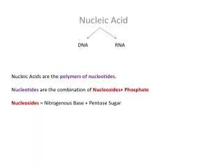
Do the differences between RNA and DNA matter? Yes! • DNA has deoxythymidine, RNA has uridine: • cytidine spontaneously degrades to uridine • dC spontaneously degrades to dU • The only dU found in DNA is there because of degradation: dT goes with dA • So when a cell finds dU in its DNA, it knows it should replace it with dC or else synthesize dG opposite the dU instead of dA Biochemistry: Nucleic Acid Chem&Struct
Ribose vs. deoxyribose • Presence of -OH on 2’ position makes the 3’ position in RNA more susceptible to nonenzymatic cleavage than the 3’ in DNA • The ribose vs. deoxyribose distinction also influences enzymatic degradation of nucleic acids • I can carry DNA in my shirt pocket, but not RNA Biochemistry: Nucleic Acid Chem&Struct
Backbone hydrolysis of nucleic acids in base(fig. 10.29) • Nonenzymatic hydrolysis in base occurs with RNA but not DNA, as just mentioned • Reason: in base, RNA can form a specific 5-membered cyclic structure involving both 3’ and 2’ oxygens • When this reopens, the backbone is cleaved and you’re left with a mixture of 2’- and 3’-NMPs Biochemistry: Nucleic Acid Chem&Struct
Enzymatic cleavage of oligo- and polynucleotides • Enzymes are phosphodiesterases • Could happen on either side of the P • 3’ cleavage is a-site; 5’ is b-site. • Endonucleases cleave somewhere on the interior of an oligo- or polynucleotide • Exonucleases cleave off the terminal nucleotide Biochemistry: Nucleic Acid Chem&Struct
An a-specific exonuclease Biochemistry: Nucleic Acid Chem&Struct
A b-specific exonuclease Biochemistry: Nucleic Acid Chem&Struct
Specificity in nucleases • Some cleave only RNA, others only DNA, some both • Often a preference for a specific base or even a particular 4-8 nucleotide sequence (restriction endonucleases) • These can be used as lab tools, but they evolved for internal reasons Biochemistry: Nucleic Acid Chem&Struct
Variety of nucleases Biochemistry: Nucleic Acid Chem&Struct
Restriction endonucleases • Evolve in bacteria as antiviral tools • “Restriction” because they restrict the incorporation of foreign DNA into the bacterial chromosome • Recognize and bind to specific palindromic DNA sequences and cleave them • Self-cleavage avoided by methylation • Types I, II, III: II is most important • I and III have inherent methylase activity; II has methylase activity in an attendant enzyme Biochemistry: Nucleic Acid Chem&Struct
What do we mean by palindromic? • In ordinary language, it means a phrase that reads the same forward and back: • Madam, I’m Adam. (Genesis 3:20) • Eve, man, am Eve. • Able was I ere I saw Elba. (Napoleon) • A man, a plan, a canal: Panama! (T. Roosevelt) • With DNA it means the double-stranded sequence is identical on both strands Biochemistry: Nucleic Acid Chem&Struct
Quirky math question to ponder • Numbers can be palindromic: • 484, 1331, 727, 595… • Some numbers that are palindromic have squares that are palindromic… • 222 = 484, 1212 = 14641, . . . • Question: if a number is perfect square and a palindrome, is its square root a palindrome? (answer will be given orally) Biochemistry: Nucleic Acid Chem&Struct
Palindromic DNA • G-A-A-T-T-C • Single strand isn’t symmetric: but the combination with the complementary strand is: • G-A-A-T-T-CC-T-T-A-A-G • These kinds of sequences are the recognition sites for restriction endonucleases. This particular hexanucleotide is the recognition sequence for EcoRI. Biochemistry: Nucleic Acid Chem&Struct
Cleavage by restriction endonucleases • Breaks can be cohesive (if they’re off-center within the sequence) or non-cohesive (blunt) (if they’re at the center) • EcoRI leaves staggered 5’-termini: cleaves between initial G and A • PstI cleaves CTGCAG between A and G, so it leaves staggered 3’-termini • BalI cleaves TGGCCA in the middle: blunt! Biochemistry: Nucleic Acid Chem&Struct
iClicker question: • Which of the following is a potential restriction site? • (a) ACTTCA • (b) AGCGCT • (c) TGGCCT • (d) AACCGG • (e) none of the above. Biochemistry: Nucleic Acid Chem&Struct
Example for EcoRI • 5’-N-N-N-N-G-A-A-T-T-C-N-N-N-N-3’3’-N-N-N-N-C-T-T-A-A-G-N-N-N-N-5’ • Cleaves G-A on top, A-G on bottom: • 5’-N-N-N-N-GA-A-T-T-C-N-N-N-N-3’3’-N-N-N-N-C-T-T-A-AG-N-N-N-N-5’ • Protruding 5’ ends:5’-N-N-N-N-GA-A-T-T-C-N-N-N-N-3’3’-N-N-N-N-C-T-T-A-AG-N-N-N-N-5’ Biochemistry: Nucleic Acid Chem&Struct
How often? • 4 types of bases • So a recognition site that is 4 bases long will occur once every 44 = 256 bases on either strand, on average • 6-base site: every 46= 4096 bases, which is roughly one gene’s worth Biochemistry: Nucleic Acid Chem&Struct
EcoRI structure • Dimeric structure enables recognition of palindromic sequence • sandwich in each monomer EcoRI pre-recognition complex PDB 1CL8 57 kDa dimer + DNA Biochemistry: Nucleic Acid Chem&Struct
Methylases HhaI methyltransferasePDB 1SVU2.66Å; 72 kDa dimer • A typical bacterium protects its own DNA against cleavage by its restriction endonucleases by methylating a base in the restriction site • Methylating agent is generally S-adenosylmethionine Structure courtesy steve.gb.com Biochemistry: Nucleic Acid Chem&Struct
Use of restriction enzymes • Nature made these to protect bacteria; we use them to cleave DNA in analyzable ways • Similar to proteolytic digestion of proteins • Having a variety of nucleases means we can get fragments in multiple ways • We can amplify our DNA first • Can also be used in synthesis of inserts that we can incorporate into plasmids that enable us to make appropriate DNA molecules in bacteria Biochemistry: Nucleic Acid Chem&Struct
Sanger dideoxy method • Incorporates DNA replication as an analytical tool for determining sequence • Uses short primer that attaches to the 3’ end of the ssDNA, after which a specially engineered DNA polymerase • Each vial includes one dideoxyXTP and 3 ordinary dXTPs; the dideoxyXTP will be incorporated but will halt synthesis because the 3’ position is blocked. • See figs. 11.3 & 11.4 for how these are read out Biochemistry: Nucleic Acid Chem&Struct
Automating dideoxy sequencing • Laser fluorescence detection allows for primer identification in real time • An automated sequencing machine can handle 4500 bases/hour • That’s one of the technologies that has made large-scale sequencing projects like the human genome project possible Biochemistry: Nucleic Acid Chem&Struct
DNA secondary structures • If double-stranded DNA were simply a straight-legged ladder: • Base pairs would be 0.6 nm apart • Watson-Crick base-pairs have very uniform dimensions because the H-bonds are fixed lengths • But water could get to the apolar bases • So, in fact, the ladder gets twisted into a helix. • The most common helix is B-DNA, but there are others. B-DNA’s properties include: • Sugar-sugar distance is still 0.6 nm • Helix repeats itself every 3.4 nm, i.e. 10 bp Biochemistry: Nucleic Acid Chem&Struct
Properties of B-DNA • Spacing between base-pairs along helix axis = 0.34 nm • 10 base-pairs per full turn • So: 3.4 nm per full turn is pitch length • Major and minor grooves, as discussed earlier • Base-pair plane is almost perpendicular to helix axis Biochemistry: Nucleic Acid Chem&Struct
Major groove in B-DNA • H-bond between adenine NH2 and thymine ring C=O • H-bond between cytosine amine and guanine ring C=O • Wide, not very deep Biochemistry: Nucleic Acid Chem&Struct
Minor groove in B-DNA • H-bond between adenine ring N and thymine ring NH • H-bond between guanine amine and cytosine ring C=O • Narrow but deep Biochemistry: Nucleic Acid Chem&Struct
Cartoon of AT pair in B-DNA Biochemistry: Nucleic Acid Chem&Struct
Cartoon of CG pair in B-DNA Biochemistry: Nucleic Acid Chem&Struct
What holds duplex B-DNA together? • H-bonds (but just barely) • Electrostatics: Mg2+ –PO4-2 • van der Waals interactions • - interactions in bases • Solvent exclusion • Recognize role of grooves in defining DNA-protein interactions Biochemistry: Nucleic Acid Chem&Struct
Helical twist (fig. 11.9a) • Rotation about the backbone axis • Successive base-pairs rotated with respect to each other by ~ 32º Biochemistry: Nucleic Acid Chem&Struct
Propeller twist • Improves overlap of hydrophobic surfaces • Makes it harder for water to contact the less hydrophilic parts of the molecule Biochemistry: Nucleic Acid Chem&Struct
A-DNA (figs. 11.10) • In low humidity this forms naturally • Not likely in cellular duplex DNA, but it does form in duplex RNA and DNA-RNA hybrids because the 2’-OH gets in the way of B-RNA • Broader • 2.46 nm per full turn • 11 bp to complete a turn • Base-pairs are not perpendicular to helix axis:tilted 19º from perpendicular Biochemistry: Nucleic Acid Chem&Struct
Z-DNA (figs. 11.10) • Forms in alternating Py-Pu sequences and occasionally in PyPuPuPyPyPu, especially if C’s are methylated • Left-handed helix rather than right • Bases zigzag across the groove Biochemistry: Nucleic Acid Chem&Struct
Getting from B to Z • Can be accomplished without breaking bonds • … even though purines have their glycosidic bonds flipped (anti -> syn) and the pyrimidines are flipped altogether! Biochemistry: Nucleic Acid Chem&Struct
DNA is dynamic • Don’t think of these diagrams as static • The H-bonds stretch and the torsions allow some rotations, so the ropes can form roughly spherical shapes when not constrained by histones • Shape is sequence-dependent, which influences protein-DNA interactions Biochemistry: Nucleic Acid Chem&Struct