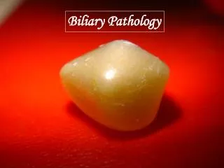
Biliary Pathology
Biliary Pathology. Liver. Biliary tree. Gallbladder. RPC. Acute cholecystitis Empyema of gallbladder Mucoceal of gallbladder. Cholangitis. Pancreatitis. Biliary Patholgy. History and Examination Previous biliary surgery Previous history of biliary pathology Epigastic / RUQ pain

Biliary Pathology
E N D
Presentation Transcript
Liver Biliary tree Gallbladder
RPC Acute cholecystitis Empyema of gallbladder Mucoceal of gallbladder Cholangitis Pancreatitis
Biliary Patholgy • History and Examination • Previous biliary surgery • Previous history of biliary pathology • Epigastic / RUQ pain • Duration of pain • Associate symptoms / signs • E.g. Tea color urine, jaundice, stool color… • Vital signs • Pulse, resp rate, blood pressure, temperature • Toxic or not ?? • X rays • Abdominal supine x ray • Gallstone • Aerobilia • CXR --- right side pleural effusion • Blood test • WCC, LFT, amylase ECG
Acute cholecystitis • Blockage of cystic duct • Tissue oedema …. Ischaemia … infection • Ultrasound abdomen • Thickened gallbladder wall • Sonographic murphy’s sign • Pericholecystic fluid • Gallstone • Conservative Vs Early Lap +/- open cholecystectomy Vs Cholecystostomy • Medical / GA risk of the patient
Acute Cholecystitis • Ultrasound abdomen • Thickened gallbladder wall • Pericholecystic Fluid • Sonographic murphy sign • Also look at: • Free fluid • CBD size / CBD stone • Gallstones
Acute Cholecystitis • Early Vs Delay Cholecystectomy • Medical risk too high for GA operation • Precutaneous Cholecystostomy • Decompress the gallbladder • Drain infected bile / pus • Still require antibiotic
Cholecystostomy • Decompress the gallbladder • Observe for improvement • Continue deterioration • ? Perforated gallbladder – urgent operation • Tube blocked • Drain content and amount • Bile production approx 500mls per day • Mucous only – blocked cystic duct • Lots of bile (500mls or above) – CBD stone • Check cholecystogram • Tube position • Position of stone in gallbladder (cystic duct still obstructed ?) • Cholangiogram (any CBD stone… drop stone) • Definitive Rx plan • Lap cholecystectomy • Conservative Rx / Removal of tube
Cholangitis Cholangitis
Fever RUQ pain Jaundice +/- Confusion Biliary Decompression Need to check clotting profile Vit K IV 10mg daily Antibiotics Definitive Mx Aim to remove causative factors Cholangitis Charcot’s triad • Assess patient clinical status • Vital parameters • Resuscitation Biliary decompression Ultra-urgent Vs urgent/early
Biliary decompression • ERCP • Endoscopic retrograde cholangiopancreatogram • PTBD • Percutaneous transhepatic biliary drainage • ECBD • Exploration of common bile duct
ERCP with previous B2 gastrectomy Definitely higher risk of perforation
Cholangitis • What if ERCP failed ?? • Anatomical problem • Technical difficulties • Patient condition poor • Respiratory problem • Severe sepsis / MOF
Percutaneous Transhepatic Biliary Drainage External PTBD
Percutaneous Transhepatic Biliary Drainage • Under LA, interventional radiological procedure • Risks • Contrast allergies, Local anaesthetic agents allergies • Liver laceration, bleeding, traumatic tapping • NOT suitable in patient with marked ascites • Tube complications • Kinking, blockage, dislodgement • Patient usually spike high fever after procedure • Need injection of contrast into biliary tree --- bacteraemia
Percutaneous Transhepatic Biliary Drainage • External PTBD • Right or left system ? • Usually communicable except in case of tumour • Usually external PTBD at initial stage • Short procedure time, less manipulation • Aim is to decompress the biliary system • Internal – external PTBD • Aim to direct bile into duodenum • Need better cholangiogram first (therefore wait till 1 wk after inflammation settle and edema subside) • Lost of fluid, electrolytes • Able to disconnect the bed side bag • Allow future ERCP (two hand technique) if initial ERCP failed due to difficult cannulation
ERCP two hand technique Internal – external PTBD ERCP
Percutaneous Transhepatic Biliary Drainage • Monitor patient progress • Vital signs • Clinical signs • Drainage amount and content • Suspect blockage revision? • Definitive Management Plan • Surgery • Trans PTBD dilatation and removal of CBD stone
Remember to use correct antibiotic Send for Timing of biliary decompression is crucial Cholangitis can be quickly lethal especially in elderly Need accurate clinical assessment and appropriate treatment
ERCP of a case of RPC Stones
RPC • Appropriate antibiotic • According to previous bile culture • Depends on the disease distribution • Peripheral and / or Central • Decompression of biliary system • Definitive Treatment • Exploration of common bile duct +/- drainage procedure • Choledochoduodenostomy • Choledochojejunostomy • Liver segmentectomy
RPC • Before definitive procedure • Need to correctly assess • Biliary drainage and function of the liver • Accurate assessment of the distribution of the disease • CT abdomen • EHIDA scan
RPC • Definitive treatments Vs conservative Plan • Need to BALANCE the risk of OT and the problems of disease process • Disease process • Recurrent attack • Liver abscess formation • Cholangiocarcinoma