Biliary System
Biliary System. Objectives. At the end of the lecture, the student should be able to describe the: Location, surface anatomy, parts, relations & peritoneal reflection of the gall bladder. Blood supply, nerve supply and lymphatic drainage of gall bladder.


Biliary System
E N D
Presentation Transcript
Biliary System Dr. Zeenat & Dr. Vohra
Objectives At the end of the lecture, the student should be able to describe the: • Location, surface anatomy, parts, relations & peritoneal reflection of the gall bladder. • Blood supply, nerve supply and lymphatic drainage of gall bladder. • Course of each of common hepatic, cystic and common bile duct.
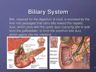
Biliary System The biliary system consists of the liver, gallbladder & bile ducts that are involved in the production, storage & transportation of bile. Bile is secreted by the liver cells at a constant rate of about 40 ml per hour. When digestion is not taking place, the bile is stored and concentrated in the gallbladder; later, it is delivered to the duodenum.
The Bile Ducts The bile ducts consist of: • Bile canaliculi • Interlobular ducts • Intrahepatic ducts • Right and left hepatic ducts • Common hepatic duct • Gallbladder & Cystic duct • Common bile duct (Bile duct)
The Bile Ducts • The liver cells secrete bile • The bile canaliculicarry this bile to the interlobular ducts. • The interlobular ducts join one another to form progressively larger ducts and, eventually, at the portahepatis form the right and left hepatic ducts.
The right hepatic duct drains the right lobe of the liver • The left hepatic duct drains the left lobe, the caudate lobe, & quadrate lobe. • After a short course, the hepatic ducts unite to form the common hepatic duct • The common hepatic duct is about 1.5 in. (4 cm) long • It descends within the free margin of the lesser omentum • It is joined on the right side by the cystic duct from the gallbladder to form the common bile duct
Common Bile Duct (Bile Duct) • The common bile duct is about 3 inches (8 cm) long. • course: • First it lies in the free margin of lesser omentum along with portal vein and hepatic artery. • Then it runs behind the first part of the duodenum. • Then it lies in a groove on the posterior surface of the head of the pancreas. Here, the bile duct comes into contact with the main pancreatic duct
Common Bile Duct Sphincter of Oddi The terminal parts of both ducts and the ampulla are surrounded by circular muscle, known as the sphincter of the hepatopancreatic ampulla (sphincter of Oddi). • The bile duct ends below by piercing the medial wall of the second part of the duodenum about halfway down its length. • It is usually joined by the main pancreatic duct, and together they open into a small ampulla in the duodenal wall, called the hepatopancreaticampulla (ampulla of Vater). The ampulla opens into the lumen of the duodenum by means of a small papilla, the major duodenal papilla. • Occasionally, the bile and pancreatic ducts open separately into the duodenum
Gallbladder • A pear-shaped sac lying on the undersurface of the liver. • It has a capacity of 30 to 50 ml and stores bile, which it concentrates by absorbing water. • The gallbladder is divided into the fundus, body, and neck. • The fundus is rounded and projects below the inferior margin of the liver, where it comes in contact with the anterior abdominal wall at the level of the tip of the ninth right costal cartilage. • The body lies in contact with the visceral surface of the liver and is directed upward, backward, and to the left. • The neck becomes continuous with the cystic duct, which turns into the lesser omentum, joins the common hepatic duct, to form the bile duct
Gallbladder • Relations • Anteriorly: The anterior abdominal wall and the inferior surface of the liver • Posteriorly: The transverse colon and the first and second parts of the duodenum • Function of the Gallbladder • When digestion is not taking place, the sphincter of Oddi remains closed and bile accumulates in the gallbladder. The gallbladder concentrates bile; stores bile; selectively absorbs bile salts, keeps the bile acid; excretes cholesterol; and secretes mucus. To aid in these functions, the mucous membrane is thrown into permanent folds that unite with each other, giving the surface a honeycombed appearance. • The peritoneum completely surrounds the fundusof the gallbladder and binds the body and neck to the visceral surface of the liver.
Nerve Supply • Sympathetic and parasympathetic vagal fibers form the celiac plexus. The gallbladder contracts in response to the hormone cholecystokinin, which is produced by the mucous membrane of the duodenum on the arrival of fatty food from the stomach. • Lymph Drainage • The lymph drains into a cystic lymph node situated near the neck of the gallbladder. From here, the lymph vessels pass to the hepatic nodes along the course of the hepatic artery and then to the celiac nodes. Blood Supply The cystic artery isa branch of the right hepatic artery. The cystic vein drains directly into the portal vein. Several very small arteries and veins also run between the liver and gallbladder.
Cystic Duct • The mucous membrane of the cystic duct is raised to form a spiral fold that is continuous with a similar fold in the neck of the gallbladder. The fold is commonly known as the “spiral valve.” The function of the spiral valve is to keep the lumen constantly open. The cystic duct is about 1.5 in. (3.8 cm) long and connects the neck of the gallbladder to the common hepatic duct to form the bile duct. It usually is somewhat S-shaped and descends for a variable distance in the right free margin of the lesser omentum.