Download
1 / 24
260 likes | 331 Vues
Panoramic imaging, also known as pantomography, provides a single tomographic image of facial structures including dental arches and supporting structures. Learn about indications, advantages, disadvantages, rotation centers, focal trough, errors, and patient positioning important for obtaining clear diagnostic images.

E N D
Panoramic Radiograph ا.د. امل رؤوف
Panoramic imaging ( also called pantomgraphy)is a technique for producing a single tomographic image of the facial structures that includes both the maxillary and mandibular dental arches and their supporting structures .It is based on the principle of the reciprocal movement of an x- ray source and image receptor around a central point or plane, called the image layer, in which the object of interest is located. Objects in front of or behind this image layer are not clearly captured because of their movement relative to the centers of rotation of the receptor and x- ray source .
Indications of panoramic image. • 1.Prior to dental surgery under general anesthesia. • 2. Assessment of third molars. • 3.Fractures of all part of the mandible. • 4.As part of an orthodontic assessment. • 5.Antral diseases of the articular surfaces of the TMJ. • 6.Vertical alveolar bone height as part of pre implants planning. • 7.To assess bony lesion or an unerupted tooth.
Advantages of panoramic images • 1- Broad coverage of the facial bones and teeth. • 2-Low patient radiation dose • 3- Convenience of the examination for the patient • 4- Use in patient unable to open their mouths • 5- Short time required to make a panoramic image ,usually in the range of 3 to 4 minutes (this include the time necessary for positioning the patient and the actual exposure cycle) • 6- Patients readily understand panoramic films ,thus they are also a useful visual aid in patient education and case presentation
Disadvantage of panoramic image • 1-Soft tissue shadows can overlie the required hard tissue structures • 2- Magnification and distortion of the final image • 3- The use of indirect action film and intensifying screens results in some loss of image quality • 4- The technique is not suitable for children under 5 years or on some patients because of the length of exposure cycle
Rotation center • In panoramic image , the film or cassette carrier and x- ray tubehead are connected and rotate around the patient during exposure . • The axis, which the cassette carrier and x- ray tubehead rotate is termed arotation center.
One of the following three basic rotation centers which is used in panoramic x- ray machines are : • - The double center rotation • - The triple center rotation • -The moving center rotation • In all the cases , the center of rotation changes as the film and tubehead rotate around the patient. This rotational change allows the image layer to conform the elliptical shape of the dental arches .
The focal trough • In panoramic radiography , focal trough or (image layer) is a theoretical concept used to determine where the dental arches must be positioned to achieve the clearest image. • The focal trough can be defined as a three- dimentional curved zone in which structures are clearly demonstrated on the panoramic image .
The structures seen on the panoramic image are primarily those located within the image layer. Objects outside the image layer are blurred , magnified , or reduced in size and are sometimes distorted to the extent of not being recognizable .
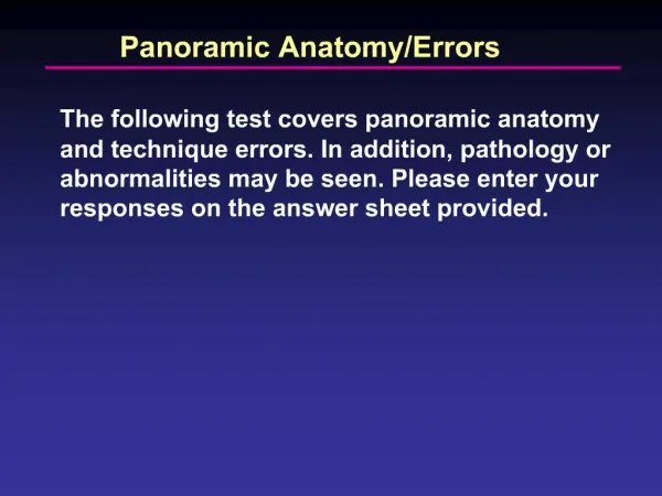
Errors in panoramic image • Proper patient preparation is critical in obtaning a diagnostic panoramic film. Two of the more common patient preparation errors include :
Ghost images: if all metallic or radiodense objects ( e.g. eyeglasses,earrings, necklace, hairpins, removable partial dentures, • orthodontic retainers and others )are not removed befored the exposure of a panoramic film, a ghost image results that obscures diagnostic information .
Lead apron arifact: if the lead apron is incorrectly placed , or if the lead apron with athyroid collar is used during the exposue of the panoramic film, a radiopaque cone-shaped artifact results that obsures diagnostic information.
Patient positioningis of critical importance during exposure of panoramic film , some errors may ocure during patient positioning for panoramic image, likes:
Positioning of the lips and tongue: if the patient,s lips are not closed on the bite -block during the exposure of the film , a dark radolucent shadow results that obscures the anterior teeth .
If the tongue is not in contact with the palate during the exposure of the film , a dark radiolucent shadow result that obscures of the maxillary teeth.
Positioning of the teeth-anterior to the focal trough: if the patient,s anterior teeth are not positioned in the focal trough indicated by the groove in the bite -block , the teeth appear blurred .If thepatient,s teeth are posithioned too far forward on the bite -block or anterior to the focal trough, the anterior teeth appear skinny and out of the radiograph
Posithioning of the teeth - posterior to the focal trough: if the patient,s anterior teeth are positioned too far back on the groove of the bite - block or posterior to the focal trough , the anterior teeth appear fat