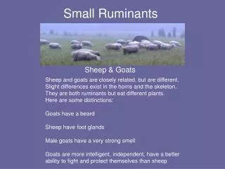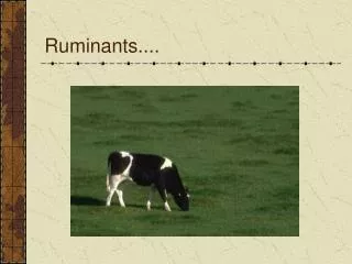
Ruminants Anatomy
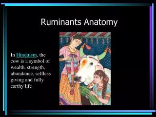
Ruminants Anatomy . In Hinduism , the cow is a symbol of wealth, strength, abundance, selfless giving and fully earthy life. Objectives – Chapter 10. Know and understand the zoological classification of the species. Know and be able to proficiently use terminology associated with the species.


Ruminants Anatomy
E N D
Presentation Transcript
Ruminants Anatomy In Hinduism, the cow is a symbol of wealth, strength, abundance, selfless giving and fully earthy life
Objectives – Chapter 10 Know and understand the zoological classification of the species. Know and be able to proficiently use terminology associated with the species. Know normal physiological data for the species, and be able to identify those that are abnormal. Identify and know the uses of common instruments relevant to the species. Describe prominent anatomical or physiological properties of the species. Identify and describe characteristics of common breeds. Describe normal living environments and husbandry needs of the species. Understand and describe specific reproductive practices of the species. Understand specific nutritional requirements of the species.
Zoological Classification Kingdom Phylum Class Order Family Genus Species Animal Chordata Mammalia Artiodactyla Bovidae Bos Tarus or Indicus
Terminology Cow: Mature female Bull: Mature male Steer: Castrated male Heifer: Immature female Calf: Neonate Heifer calf: Neonate female less than one year of age. Can be called first, second, third or fourth calf heifers. Bull calf: Neonate male younger than 1 year of age Calving: The act of parturition
Bovine Skeleton 7, 13, 6, 5 (fused), 18-20: Olecranon; Ligamentum nuchae
Types - bones vertebrae and certain facial bones humerus, radius, femur, tibia, metacarpals, and metatarsals patella, and proximal and distal sesamoid bones of the digits. sternum, ribs, scapula, and certain skull bones carpal and tarsal bones
Cow and Sheep Skull • The brain is situated within the cranium - a box-like posterior part of the skull. • The brain is connected to the spinal cord through a large hole, the foramen magnum. • The foramen magnum is flanked by two large knobs or occipital condyles that form a joint with the first cervical vertebra of the neck. • Sinuses or spaces are present between the inner and outer cranial walls.
Nasal sinus: MCF Digital section of head with removal of nasal septum, showing accumulated fibrinous secretions lodged in the anterior portions of the left nasal turbinates (arrow).
Coronoid process is located medially to the zygomatic arch Process allows muscle leverage to be exerted onto the mandibleMandibular condyle: joint between the skull and the lowerIn cattle and sheep, the mandibular condyle is relatively flat and allows considerable movement in a horizontal plane. Lateral movement is important in animals whose teeth work with a grinding action.
Ruminants such as cattle, sheep and goats are herbivores with a unique digestive system. • A prominent feature of ruminant dental anatomy is that they lack upper incisors, having instead a "dental pad", as shown in the image to the right of a goat.
Dental Formulae 0 0 3 3 1 3 0 0 3 3 3 1 3 3 The dental anatomy of all ruminants is similar • In the dental formulae shown above, cattle are depicted as having 3 incisors and 1 canine tooth. Some authors prefer to state that they have 4 incisors, with the canine tooth refered to as the fourth or cornerincisor. • Llamas* Deciduous =10 Permanent =16
Llama and Alpacas Maxilla and mandible, llama. Maxillary teeth shown are the third incisor and canine. Mandibular teeth shown are I1-I4. The llama fighting teeth are the upper third incisors, upper canines, and lower fourth incisors (six total teeth). The fighting teeth Courtesy of Dr. Bradford B. Smith and Dr. Karen I. Timm
Tooth Eruption • CD
Maxillary Arcade • Note the lack of incisors
Mandibular Arcade(Lateral view) The wide gap that separates the incisors (or dental pad on the maxilla) from the premolars is called the diastema.
The vertebral column or backbone is the main axis of the skeleton and it protects the spinal cord. • The spinal cord is located in a neural canal formed by a long series of neural arches, each contributed by a different vertebra. • The neural arch of each vertebra is supported on the body or centrum of the vertebra. In some types of vertebrae, the neural arch extends dorsally as a prominent spine that may be called a dorsal spine, a neural spine or a spinous process.
types of vertebrae • NAME--------REGION---------BEEF--------------LAMB • Cervical--------Neck------------------7-----------------------7 • Thoracic-------Ribcage---------------13---------------------13 to 14 • Lumbar--------Loin--------------------6----------------------6 to 7 • Sacral ---------Sirloin------------------5----------------------4 • Caudal---------Tail--------------------18 to 20--------------16 to 18
assist the extensor muscles of the head and neck, extending from the occipital bone to the spinous processes of the thoracic vertebrae Plan of neck in beef, showing:1, ligamentum nuch; 2, atlas; and 3, axis. The ligamentum nuchae is pale yellow • The first cervical vertebra, the atlas, articulates with the skull and is greatly modified in shape to form a joint that enables the animal to nod its head up and down. • Rotation or twisting of the head occurs from the joint between the atlas and the next cervical vertebra, the axis. • The ligamentum nuchae is a very strong elastic ligament in the dorsal midline of the neck, and it relieves the animal of the weight of its head. Were it not for the ligamentum nuchae, the head of the standing animal would droop between its forelimbs
Contagious Bovine Pleuropneumoniae Exposed Atlanto-Occipital joint with severe thickening of the meninges and fibrin deposits
Ribcage • The cage formed by thoracic vertebrae, ribs and sternum is an essential component of the respiratory system. • Thoracic vertebrae are distinguished by their tall dorsal spines, many of which point towards the hindquarter and are known as the feather bones.
The structure of the ribcage is rather variable in lamb carcasses BEEF----------LAMB Total pairs of ribs-------------13--------------13 to 14 Pairs of sternal ribs-----------8----------------8 Pairs of asternal ribs----------5---------------5 to 6 Number of sternebrae--------7---------------6 to7
Pelvis • The pelvis and the sacrum form a ring of bone completed ventrally by the pubes. • The left pubis is separated from the right pubis by fibrocartilage which, at parturition, may soften to allow movement between the bones of the pelvis. • The pubes are separated when carcasses are split into left and right sides in the abattoir. V Plan of the pelvis in a hanging beef carcass showing:1, lesser sciatic notch; 2, ischiatic spine; 3, greater sciatic notch; 4, psoas tubercle; 5, obturator foramen; 6, symphysis pubis;7, ischium; and 8, ilium.
Pubic The tuber coxae forms the basis of the point of the hip (hooks) Another plan of the both sides of the pelvis in a hanging carcass showing: 1, tuber coxae; 2, acetabulum; 3, acetabular ramus of ischium; 4, tuber ischii; 5, symphysis pubis; 6, ilium; 7, pubis; and 8, ischium
OS COXAE - PELVIS The pelvic girdle comprised of the illium, ishium, and pubis. This is the largest of the the flat bones
Ilium – Ischium - Pubis • Smallest of the three parts of the pelvic girdle • The largest and most anterior of the three parts of the pelvic girdle • Hip bone/ Pin bone
Aitch bone – Body of shaft of Ischium • The aitch bone is curved in steer and bull carcasses, is moderately curved in heifers, but is straight in cow carcasses
Forelimb skeleton -Scapula • The scapula is not fused to the vertebral column (like the pelvis in the hindlimb), and this allows muscles that hold the scapula to the ribcage to function as shock absorbers during locomotion. • The scapula has a distal socket joint for the next bone in the forelimb, the humerus. • This socket of the glenohumeral joint is called the glenoid cavity . • The glenoid cavity is wide and shallow, unlike the ball and socket joint in the hindlimb which is narrow and deep.
ACROMION • On the lateral face of the scapula is a prominent ridge of bone called the spine of the scapula. • In beef (OX) carcasses, the scapular spine is extended distally as a prominent acromion process.
Humerus – “Arm bone / clod bone” • Proceeding distally down the forelimb, the bone that articulates with the scapula is the humerus. • Proximally, the humerus has a relatively flat knob or head to fit into the glenoid cavity of the scapula. Two well defined condyles on the distal end of the humerus contribute to the hinge joint at the elbow.
Radius & Ulna: (‘Foreshank bone’) Beef shankbones showing: 1, distal end of humerus; 2, olecranon fossa; 3, olecranon process;, 4,radius; 5, ulna; and 6, carpal bones. • The radius is joined to the ulna and is the shorter and more anterior bone of the pair
Femur – ‘Round bone or leg bone’ • The proximal bone of the hindlimb is the femur or round bone. The articular head of the femur is deeply rounded and it bears a round ligament that holds it into the acetabulum. • Another distinctive feature of the femur is the broad groove between the two trochlear ridges located distally. The patella or knee cap slides in this groove
Tibia – ‘hind shank – hock bone’ • In beef and lamb carcasses there is a single major bone, the tibia or shank bone, located distally to the femur. • Tibia and fibula 1, medial condyle, 2, lateral condyle; 3, tibia, and 4, fibula.
Foot, Digits, Claws and Dewclaws foot (Dyce) digits or toes dewclaw (hoof only) fetlock jt. pastern jt. coffin jt. bulb (heel) sole wall claws (hoof)
Common and Lateral Digital Extensor Tendons common digital extensor: Note: “just like” the horse, but double because 2 digits. medial head lateral digital extensor lateral head Note: three palpable extensor tendons, rather than two as in the horse. Dorsal view: IV III L M
Cloven (split) hoof: Cattle/ goat/ sheep 2 digits: III and IV with 3 phalanges. Digits II and V: vestiges
Erosion on the foot caused by FMD or Vesicular Stomatitis which are grossly indistinguishable from one another.
References • http://w3.vet.cornell.edu/virtualvet/bovine/tissue_lesions.aspx?Tis=37 • http://bovine.unl.edu/bovine3D/eng/nIntro.jsp • http://studentvet.wordpress.com/2010/07/29/bovine-forelimb/#Humerus • McBride Douglas, Learning Veterinary terminology, 2002 • http://vetmed.illinois.edu/courses/imaging_anatomy/bovine/hindlimb/foot/ex01/ex01.html • K Holtgrew-Bohling , Large Animal Clinical Procedures for Veterinary Technicians, 2nd Edition, Mosby, 2012 • www.vet.k-state.edu/depts/ap/faculty/klimek/.../B-P248-268.ppt