Chronic Renal Obstruction and Infectious Complications in a 60-Year-Old Female Patient
A 60-year-old female presents with bilateral flank pain, fevers, chills, nausea, and vomiting. She has a documented urinary tract infection and a history of renal stones. Imaging reveals bilateral calcific densities and pronounced renal changes in the right kidney, including enlarged cystic structures and associated stones leading to chronic obstruction. The left kidney shows a non-obstructive cyst. A diuretic renal scan is planned to assess renal function and obstruction status. Possible diagnoses include obstructive nephropathy and recurrent urinary tract infection.

Chronic Renal Obstruction and Infectious Complications in a 60-Year-Old Female Patient
E N D
Presentation Transcript
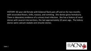
HISTORY: 60 year-old female with bilateral flank pain off and on for two months with associated fevers, chills, nausea, and vomiting. She denies gross hematuria. There is laboratory evidence of a urinary tract infection. She has a history of renal stones with several interventions, the last approximately 10 years ago. The kidney stones were calcium oxalate and struvite stones. Slide 1
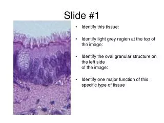
Describe the findings on the scout film and noncontrast CT (hover the cursor over CT to scroll). Slide 2
Scout Image – Bilateral calcific densities overlying the renal shadows Axial Non-contrast CT: - Right kidney: Enlarged, thinning of renal cortex, multiple large communicating cystic structures (measuring water density) consistent with chronic obstruction, many with calcific stones and fragments up to 4 cm, mild hydroureter, distal ureteral stone - Left kidney: single cystic structure, nonobstructing stones, no hydronephrosis or hydroureter -Multiple surgical clips Slide 3
The diuretic renal scan requested to evaluate function and for obstruction. What information has been incorrectly provided and why? 3) What is the likely diagnosis? Slide 7