Chapter 42
Chapter 42. Circulation and Gas Exchange. 903-905, 911-915, 919-925. Concept 42.2: Coordinated cycles of heart contraction drive double circulation in mammals. The mammalian cardiovascular system meets the body’s continuous demand for O 2. Mammalian Circulation.

Chapter 42
E N D
Presentation Transcript
Chapter 42 Circulation and Gas Exchange 903-905, 911-915, 919-925
Concept 42.2: Coordinated cycles of heart contraction drive double circulation in mammals • The mammalian cardiovascular system meets the body’s continuous demand for O2
Mammalian Circulation • Blood begins its flow with the right ventricle pumping blood to the lungs • In the lungs, the blood loads O2 and unloads CO2 • Oxygen-rich blood from the lungs enters the heart at the left atrium and is pumped through the aorta to the body tissues by the left ventricle • The aorta provides blood to the heart through the coronary arteries
Blood returns to the heart through the superior vena cava (blood from head, neck, and forelimbs) and inferior vena cava (blood from trunk and hind limbs) • The superior vena cava and inferior vena cava flow into the right atrium Animation: Path of Blood Flow in Mammals
Fig. 42-6 Capillaries of head and forelimbs Superior vena cava 7 Pulmonary artery Pulmonary artery Capillaries of right lung Aorta 9 Capillaries of left lung 3 3 2 4 11 Pulmonary vein Pulmonary vein 5 1 Right atrium Left atrium 10 Right ventricle Left ventricle Inferior vena cava Aorta Capillaries of abdominal organs and hind limbs 8
The Mammalian Heart: A Closer Look • A closer look at the mammalian heart provides a better understanding of double circulation
Fig. 42-7 Pulmonary artery Aorta Pulmonary artery Right atrium Left atrium Semilunar valve Semilunar valve Atrioventricular valve Atrioventricular valve Right ventricle Left ventricle
The heart contracts and relaxes in a rhythmic cycle called the cardiac cycle • The contraction, or pumping, phase is called systole • The relaxation, or filling, phase is called diastole
Fig. 42-8-1 Semilunar valves closed AV valves open 0.4 sec Relaxation phase, blood returning from the large veins flows into the atria and ventricles through AV valves 1 Atrial and ventricular diastole Cardiac cycle
Fig. 42-8-2 Atrial systole; ventricular diastole 2 Semilunar valves closed A brief period of atria systole then forces all blood remaining in the atria into the ventricles 0.1 sec AV valves open 0.4 sec Relaxation phase, blood returning from the large veins flows into the atria and ventricles through AV valves 1 Atrial and ventricular diastole Cardiac cycle
Fig. 42-8 Atrial systole; ventricular diastole 2 Semilunar valves closed A brief period of atria systole then forces all blood remaining in the atria into the ventricles 0.1 sec Semilunar valves open AV valves open 0.4 sec 0.3 sec Relaxation phase, blood returning from the large veins flows into the atria and ventricles through AV valves 1 Atrial and ventricular diastole Ventricular systole pumps blood into the large arteries through the semilunar valves AV valves closed Cardiac cycle 3 Ventricular systole; atrial diastole
The heart rate, also called the pulse, is the number of beats per minute • The stroke volume is the amount of blood pumped in a single contraction (average ~ 70ml) • The cardiac output is the volume of blood pumped into the systemic circulation per minute and depends on both the heart rate and stroke volume • 70 ml x 72 beats/min = 5 L/min
Four valves prevent backflow of blood in the heart, made of flaps of connective tissue • The atrioventricular (AV) valves separate each atrium and ventricle • The semilunar valves control blood flow to the aorta and the pulmonary artery
The “lub-dup” sound of a heart beat is caused by the recoil of blood against the AV valves (lub) then against the semilunar (dup) valves • Backflow of blood through a defective valve causes a heart murmur
Maintaining the Heart’s Rhythmic Beat • Some cardiac muscle cells are self-excitable, meaning they contract without any signal from the nervous system
The sinoatrial (SA) node, or pacemaker, sets the rate and timing at which cardiac muscle cells contract (wall of the right atrium near where the superior vena cava enters the heart) • Impulses from the SA node travel to the atrioventricular (AV) node • At the AV node, the impulses are delayed and then travel to the Purkinje fibers that make the ventricles contract
Impulses that travel during the cardiac cycle can be recorded as an electrocardiogram (ECG or EKG)
Fig. 42-9-1 1 Pacemaker generates wave of signals to contract. SA node (pacemaker) ECG The control of heart rhythm
Fig. 42-9-2 2 Signals are delayed at AV node. AV node The control of heart rhythm
Fig. 42-9-3 3 Signals pass to heart apex. Bundle branches Heart apex The control of heart rhythm
Fig. 42-9-4 Signals spread throughout ventricles. 4 Purkinje fibers The control of heart rhythm
Fig. 42-9-5 3 1 2 Pacemaker generates wave of signals to contract. Signals are delayed at AV node. Signals pass to heart apex. Signals spread throughout ventricles. 4 SA node (pacemaker) AV node Purkinje fibers Bundle branches Heart apex ECG The control of heart rhythm
The pacemaker is influenced by nerves, hormones (epinephrine), body temperature (1 °C →↑ 10 beats per minute), and exercise
Concept 42.4: Blood components function in exchange, transport, and defense • In invertebrates with open circulation, blood (hemolymph) is not different from interstitial fluid • Blood in the circulatory systems of vertebrates is a specialized connective tissue
Blood Composition and Function • Blood consists of several kinds of cells suspended in a liquid matrix called plasma • The cellular elements occupy about 45% of the volume of blood
Fig. 42-17 Plasma 55% Constituent Major functions Cellular elements 45% Cell type Number per µL (mm3) of blood Functions Solvent for carrying other substances Water Erythrocytes (red blood cells) Transport oxygen and help transport carbon dioxide 5–6 million Ions (blood electrolytes) Sodium Potassium Calcium Magnesium Chloride Bicarbonate Osmotic balance, pH buffering, and regulation of membrane permeability Separated blood elements Leukocytes (white blood cells) Defense and immunity 5,000–10,000 Plasma proteins Albumin Osmotic balance pH buffering Lymphocyte Basophil Fibrinogen Clotting Eosinophil Immunoglobulins (antibodies) Defense Neutrophil Monocyte Substances transported by blood Nutrients (such as glucose, fatty acids, vitamins) Waste products of metabolism Respiratory gases (O2 and CO2) Hormones 250,000– 400,000 Platelets Blood clotting
Plasma • Blood plasma is about 90% water • Among its solutes are inorganic salts in the form of dissolved ions, sometimes called electrolytes • Another important class of solutes is the plasma proteins, which influence blood pH, osmotic pressure, and viscosity • Various plasma proteins function in lipid transport, immunity, and blood clotting
Cellular Elements • Suspended in blood plasma are two types of cells: • Red blood cells (erythrocytes) transport oxygen • White blood cells (leukocytes) function in defense • Platelets, a third cellular element, are fragments of cells that are involved in clotting
Erythrocytes • Red blood cells, or erythrocytes, are by far the most numerous blood cells • They transport oxygen throughout the body • They contain hemoglobin, the iron-containing protein that transports oxygen
Leukocytes • There are five major types of white blood cells, or leukocytes: monocytes, neutrophils, basophils, eosinophils, and lymphocytes • They function in defense by phagocytizing bacteria and debris or by producing antibodies • They are found both in and outside of the circulatory system
Platelets • Platelets are fragments of cells and function in blood clotting
Blood Clotting • When the endothelium of a blood vessel is damaged, the clotting mechanism begins • A cascade of complex reactions converts fibrinogen to fibrin, forming a clot • A blood clot formed within a blood vessel is called a thrombus and can block blood flow
Fig. 42-18-1 Collagen fibers Platelet plug Platelet releases chemicals that make nearby platelets sticky
Fig. 42-18-2 Collagen fibers Platelet plug Platelet releases chemicals that make nearby platelets sticky Clotting factors from: Platelets Damaged cells Plasma (factors include calcium, vitamin K)
Fig. 42-18-3 Collagen fibers Platelet plug Platelet releases chemicals that make nearby platelets sticky Clotting factors from: Platelets Damaged cells Plasma (factors include calcium, vitamin K) Prothrombin Thrombin
Fig. 42-18-4 Red blood cell Collagen fibers Platelet plug Fibrin clot Platelet releases chemicals that make nearby platelets sticky Clotting factors from: Platelets Damaged cells Plasma (factors include calcium, vitamin K) Prothrombin Thrombin Fibrinogen Fibrin 5 µm
Stem Cells and the Replacement of Cellular Elements • The cellular elements of blood wear out and are replaced constantly throughout a person’s life • Erythrocytes, leukocytes, and platelets all develop from a common source of stem cells in the red marrow of bones • The hormone erythropoietin (EPO) stimulates erythrocyte production when oxygen delivery is low
Fig. 42-19 Stem cells (in bone marrow) Myeloid stem cells Lymphoid stem cells Lymphocytes B cells T cells Neutrophils Erythrocytes Platelets Eosinophils Basophils Monocytes
Cardiovascular Disease • Cardiovascular diseases are disorders of the heart and the blood vessels • They account for more than half the deaths in the United States
Atherosclerosis • One type of cardiovascular disease, atherosclerosis, is caused by the buildup of plaque deposits within arteries
Fig. 42-20a Smooth muscle Connective tissue Endothelium 50 µm (a) Normal artery
Fig. 42-20b Plaque (b) Partly clogged artery 250 µm
Heart Attacks and Stroke • A heart attack is the death of cardiac muscle tissue resulting from blockage of one or more coronary arteries • A stroke is the death of nervous tissue in the brain, usually resulting from rupture or blockage of arteries in the head
Treatment and Diagnosis of Cardiovascular Disease • Cholesterol is a major contributor to atherosclerosis • Low-density lipoproteins (LDLs) are associated with plaque formation; these are “bad cholesterol” • High-density lipoproteins (HDLs) reduce the deposition of cholesterol; these are “good cholesterol” • The proportion of LDL relative to HDL can be decreased by exercise, not smoking, and avoiding foods with trans fats
Statins: drugs given to people at high risk that lowers LDL levels • Aspirin: blocks the inflammatory response, help prevent the recurrence of heart attacks and stroke • C-reactive protein (CRP) produced by the liver during inflammation. High CRP in the blood is a predictor of cardiovascular disease.
Hypertension, or high blood pressure, promotes atherosclerosis and increases the risk of heart attack and stroke • Chronic high blood pressure damages the endothelium that lines the arteries, promoting plaque formation • Hypertension → when systolic pressure above 140 mm Hg or diastolic pressure above 90 mm Hg. • Hypertension can be reduced by dietary changes, exercise, and/or medication
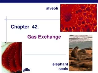
Concept 42.5: Gas exchange occurs across specialized respiratory surfaces • Gas exchange supplies oxygen for cellular respiration and disposes of carbon dioxide
Lungs • Lungs are an infolding of the body surface • The circulatory system (open or closed) transports gases between the lungs and the rest of the body • The size and complexity of lungs correlate with an animal’s metabolic rate
Mammalian Respiratory Systems: A Closer Look • A system of branching ducts conveys air to the lungs • Air inhaled through the nostrils passes through the pharynx via the larynx, trachea, bronchi, bronchioles, and alveoli, where gas exchange occurs • Exhaled air passes over the vocal cords to create sounds • Secretions called surfactants (phospholipids and proteins)coat the surface of the alveoli required to relive the surface tension in the fluid that coat their surface.
Fig. 42-24 Branch of pulmonary vein (oxygen-rich blood) Branch of pulmonary artery (oxygen-poor blood) Terminal bronchiole Nasal cavity Pharynx Larynx Alveoli (Esophagus) Left lung Trachea Right lung Bronchus Bronchiole Diaphragm Heart SEM Colorized SEM 50 µm 50 µm