Download
1 / 23
250 likes | 279 Vues
Explore the intricate world of epigenetics in cellular differentiation, identifying key mechanisms influencing gene expression and cellular programs. Understand the reversible yet stable phenotypic states governed by epigenetic modifications such as DNA methylation and histone modifications.

E N D
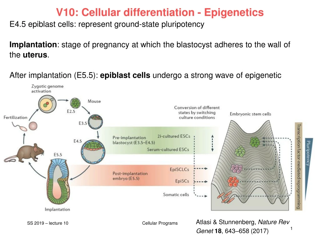
V10: Cellular differentiation - Epigenetics E4.5 epiblast cells: represent ground-state pluripotency Implantation: stage of pregnancy at which the blastocyst adheres to the wall of the uterus. After implantation (E5.5): epiblastcellsundergo a strong waveofepigeneticreprogramming. Theyarenow „primed“. Atlasi & Stunnenberg, Nature Rev Genet18, 643–658 (2017) Cellular Programs
Epigenetic mechanisms Epigeneticsreferstoalternatephenotypicstatesthatare not based on differences in genotype, andarepotentially reversible, but aregenerallystablymaintainedduringcelldivision. Examples: imprinting, twins, cancer vs. normal cells, differentiation, ... Multiple mechanismsinteracttocollectivelyestablish - alternatestatesofchromatinstructure (open – packed/condensed), - histonemodifications, • compositionofassociatedproteins (e.g. histones), • transcriptionalactivity, • activityofmicroRNAs, and - in mammals, cytosine-5 DNA methylation at CpGdinucleotides. Laird, Hum Mol Gen 14, R65 (2005) Cellular Programs
Waddington’s epigenetic landscape for embryology Waddington worked in embryology a) is a painting by John Piper that was used as the frontispiece for Waddington's book Organisers and Genes. It represents an epigenetic landscape. Developmental pathways that could be taken by each cell of the embryo are metaphorically represented by the path taken by water as it flows down the valleys. b) Later depiction of the epigenetic landscape. The ball represents a cell, and the bifurcating system of valleys represents bundles of trajectories in state space. Conrad Hal Waddington (1905 – 1975) pictures.royalsociety.org Slack, Nature Rev Genet 3, 889-895 (2002) Cellular Programs
Cytosine methylation Observation: 3-6 % of all cytosines are methylated in human DNA. This methylation occurs (almost) exclusively when cytosine is followed by a guanine base -> CpG dinucleotide. 5-methyl-cytosine SAM: S-adenosyl-methionine SAH: S-adenosyl-homocysteine Cytosine Mammalian genomes contain much fewer (only 20-25 %) of the CpG dinucleotide than is expected by the G+C content (we expect 1/16 ≈ 6% for any random dinucleotide). This is typically explained in the following way: As most CpGs serve as targets of DNA methyltransferases, they are usually methylated …. (see following page) Esteller, Nat. Rev. Gen. 8, 286 (2007) www.wikipedia.org Cellular Programs
Cytosine methylation But 5-Methylcytosine can easily deaminate to thymine. If this mutation is not repaired, the affected CpG is permanently converted to TpG (or CpA if the transition occurs on the reverse DNA strand). Hence, methylCpGs represent mutational hot spots in the genome. If such mutations occur in the germ line, they become heritable. A constant loss of CpGs over thousands of generations can explain the low frequency of this special dinucleotide in the genomes of human and mouse. 5-methyl-cytosine thymine Esteller, Nat. Rev. Gen. 8, 286 (2007) www.wikipedia.org Cellular Programs
chromatin organization affects gene expression Schematic of the reversible changes in chromatin organization that influence gene expression: genes are expressed (switched on) when the chromatin is open (active), and they are inactivated (switched off) when the chromatin is condensed (silent). White circles = unmethylated cytosines; red circles = methylated cytosines. Rodenhiser, Mann, CMAJ 174, 341 (2006) Cellular Programs
Altered DNA methylation upon cancerogenesis Genomic Imprinting: Mono-allelic expression; one allele (eitherfrom themotherorthe father) issilenced. Typically, thisis implementedby methylatingthe silenced allele. The human genome contains ca. 8% of retroviral sequences. Typically, theseare also silencedby DNA methylation. Esteller, Nat. Rev. Gen. 8, 286 (2007) Cellular Programs
Enzymes that controlDNA methylation and histone modfications These dynamicchromatinstatesarecontrolledby reversible epigeneticpatternsofDNA methylationandhistonemodifications. Enzymes involved in thisprocessinclude - DNA methyltransferases (DNMTs), - histonedeacetylases (HDACs), - histoneacetylases, - histonemethyltransferases(HMT) andthe • methyl-bindingdomainproteinMECP2 withits methyl-bindingdomain (MBD) thatbindsspecificallytome-cytosine. HP1: heterochromatinprotein 1 Rodenhiser, Mann, CMAJ 174, 341 (2006) Feinberg AP & Tycko P (2004) Nature Reviews: 143-153 Cellular Programs
DNA methylation Typically, unmethylatedclustersofCpGpairsarelocated in tissue-specific genesand in essential housekeeping genes. (House-keeping genes areinvolved in routinemaintenancerolesandareexpressed in mosttissues.) These clusters, orCpGislands, aretargetsforproteins that bind tounmethylatedCpGsandinitiategenetranscription. In contrast, methylatedCpGsaregenerallyassociatedwithsilent DNA, can block methylation-sensitive proteinsandcanbeeasilymutated. The lossof normal DNA methylationpatternsisthe bestunderstoodepigeneticcauseofdisease. In animalexperiments, theremovalof genes thatencode DNMTs islethal; in humans, overexpressionoftheseenzymeshasbeenlinked to a varietyofcancers. Rodenhiser, Mann, CMAJ 174, 341 (2006) Cellular Programs
Higher forms of methylation – Tet enzymes Unmodifiedcytosine (C) ismethylatedby DNA methyltransferases (DNMTs) at the 5 positiontobecome 5-methylcytosine (5mC). TET proteinsoxidize 5mC into 5-hydroxymethylcytosine (5hmC), a stableepigeneticmark, andsubsequentlyto 5-formylcytosine (5fC) and 5-carboxylcytosine (5caC). TET candemethylate DNA via replication-dependent (passive) orreplication-independent (active) mechanisms. Lio & Rao, Front. Immunol. (2019) CellularPrograms
Higher forms of methylation – abundance The approximate abundance of unmodified and modified cytosines in the haploid human/mouse genome. About 5% of cytosine is methylated (5mC); in most cells, the vast majority of 5mC is present at CG dinucleotides although it is low at CpG islands. 5hmC amounts to about 1-10% of 5mC (estimated at 10% here as in embryonic stem cells), while the levels of 5fC and 5caC are each about an order of magnitude lower than the previous oxidative modification. Lio & Rao, Front. Immunol. (2019) CellularPrograms
Passive DNA methylation The DNMT1/UHRF1 complex recognizes 5mC at the hemi-methylated CpG motif during DNA replication and methylates the unmodified cytosine on the newly synthesized DNA strand. However, the oxidized methylcytosines 5hmC, 5fC, and 5caC are not recognized by DNMT1/UHRF1, resulting in unmodified cytosine on the new DNA strand. Further DNA replication in the presence of continuing TET activity will result in progressive dilution of 5mC in the daughter cells. Lio & Rao, Front. Immunol. (2019) CellularPrograms
Active DNA methylation While 5hmC is stable and persists in the genome, 5fC and 5caC can be recognized and excised by thymine DNA glycosylase (TDG), and the resulting abasic sites are repaired as unmodified C by base excision repair (BER). Lio & Rao, Front. Immunol. (2019) CellularPrograms
(review V2) The histone code The DNA of eukaryotic organisms is packaged into chromatin, whose basic repeating unit is the nucleosome. A nucleosome is formed by wrapping 147 base pairs of DNA twice around an octamer of four core histones, H2A , H2B , H3 and H4 (2 copies of each one). X-ray structure of the nucleosome core particle consisting of core histones, and DNA. Top view. Side view shows two windings of DNA and two histone layers www.wikipedia.org Cellular Programs
(review V2) Post-translational modifications of histone tails The disordered histone tails comprise 25-30% of the histone mass. They extend from the compact histone multimer to provide a platform for various post-translational modifications (PTMs). These modifications affect the histones' ability to bind DNA and to other histones. This, in turn, affects gene expression. PNAS 1964;51:786 First report on PTMs of histones Strahl BD and Allis CD, 2000. Nature 403:41-45 Cellular Programs
Mode of action of histone PTMs Histone PTMs exert their effects via two main mechanisms. (1) PTMs directly influence the overall structure of chromatin, either over short or long distances. (2) PTMs regulate (either positively or negatively) the binding of effector molecules. Bannister, Kouzarides, Cell Res. (2011) 21: 381–395. Cellular Programs
PTMs of histone tails Histone acetylation and phosphorylation effectively reduce the positive charge of histones. This potentially disrupts electrostatic interactions between histones and DNA. This presumably leads to a less compact chromatin structure, thereby facilitating DNA access by protein machineries such as those involved in transcription. Histone methylation mainly occurs on the side chains of lysines and arginines. Unlike acetylation and phosphorylation, however, histone methylation does not alter the charge of the histone protein. Bannister, Kouzarides, Cell Res. (2011) 21: 381–395. By Ybs.Umich - Own work, CC BY-SA 3.0, https://commons.wikimedia.org/w/index.php?curid=31240656 Cellular Programs
Protein domains bind to modified histones Examples of proteins with domains that specifically bind to modified histones. There are more domain types recognizing lysine methylation than any other PTM. H3K4me3 – a mark associated with active transcription – is recognized by a PHD finger within the ING family of proteins (ING1-5). The ING proteins in turn recruit additional chromatin modifiers such as HATs and HDACs. Bannister, Kouzarides Cell Res. (2011) 21: 381–395. Cellular Programs
Epifactors database The database EpiFactors stores detailed and curated information about 815 proteins and 69 complexes involved in epigenetic regulation. http://epifactors.autosome.ru/protein_complexes Side view shows two windings of DNA and two histone layers Database (Oxford). 2015; 2015: bav067. Cellular Programs
Dynamics of epigenetic modifications DNA methylation is erased in the paternal and maternal genomes after fertilization and is put back on at later developmental stages. Atlasi & Stunnenberg, Nature Rev Genet18, 643–658 (2017) Cellular Programs
Events during enhancer activation / decommissioning Pioneer factors: transcription factors that can directly bind condensed chromatin. 5mC: 5-methyl-cytosine 5hmC: 5-hydroxy-methyl-cytosine Atlasi & Stunnenberg, Nature Rev Genet18, 643–658 (2017) Cellular Programs
Interplay between DNA methylation and histone modifications Bivalent chromatin are segments of DNA, bound to histone proteins, that have both repressing and activating epigenetic regulators in the same region. These regulators work to enhance or silence the expression of genes.Since these regulators work in opposition to each other, they normally interact with chromatin at different times. However, in bivalent chromatin, both types of regulators are interacting with the same domain at the same time.Bivalent chromatin domains are normally associated with promoters of transcription factor genes that are expressed at low levels.Bivalent domains have also been found to play a role in developmental regulation in pluripotent embryonic stems cells, as well as gene imprinting. Atlasi & Stunnenberg, Nature Rev Genet18, 643–658 (2017) www.wikipedia.org CellularPrograms
Paper #8 An IntrinsicEpigeneticBarrierforFunctional Axon Regeneration Yi Lan Weng, Ran An, Jessica Cassin, Jessica Joseph, Ruifa Mi, Chen Wang, Chun Zhong,Seung-Gi Jin, Gerd P. Pfeifer, Alfonso Bellacosa, Xinzhong Dong, Ahmet Hoke, Zhigang He, Hongjun Song, Guo-li Ming* Neuron 94, 337-346.e6 (2017) Paper presentation June 25, 2019 see also Scarlett J. Barker, Li-HueiTsai MethyLock: DNA DemethylationIstheEpigenetic Key to Axon Regeneration Neuron, 94, 221-223 (2017) CellularPrograms


