Download
1 / 56
800 likes | 1.12k Vues
PULMONARY HYPERTENSION. INTRODUCTION BRIEF HISTORY WHAT IS PH? WHAT CAUSES PH? SIGNS AND SYMPTOMS DIAGNOSIS OF PH TREATMENT OF PH. INTRODUCTION.

E N D
INTRODUCTION BRIEF HISTORY WHAT IS PH? WHAT CAUSES PH? SIGNS AND SYMPTOMS DIAGNOSIS OF PH TREATMENT OF PH
INTRODUCTION In the human body, there are two types of circulation that enable distribution of blood throughout the body. The portion that pumps oxygenated blood from the left side of the heart via the left ventricle to all parts of the body is known as the SYSTEMIC CIRCULATION. On the other hand, the portion that pumps deoxygenated blood from the right side of the heart via the right ventricle into the lungs to obtain oxygen is referred to as the PULMONARY CIRCULATION.
INTRODUCTION Millions of people are affected by a condition known as high blood pressure (hypertension) whereby the blood travels through the body’s arteries at a pressure higher than normal. PULMONARY HYPERTENSION is a far less common type of high blood pressure that affects specifically the arteries in the lungs. Pressures in the lung arteries are normally significantly lower than the pressures in the systemic circulation. Pulmonary hypertension occurs when the pressure in the pulmonary circulation becomes abnormally elevated. It is a serious condition that becomes progressively worse and eventually proves fatal.
INTRODUCTION An estimated of 500 – 1000 new cases are diagnosed annually. There is an incidence of about 2-3 per million per year and a prevalence of 15 per million. This disease can occur in men, women and children of all ages. However, it is most common in females between 20 and 40 years old, with twice as many cases reported in women then men. The condition is rare in children but is sometimes seen in infants born with heart defects. Pulmonary hypertension may be a primary or secondary cause of hypoxia in neonates.
H I S T O R Y • The first reported case – 1891 • E. Romberg, German doctor • published description of a patient who, at autopsy, showed • thickening of the pulmonary artery but no heart or lung disease • In 1951, 39 cases were reported by Dr. D.T. Dresdale in the United States. The illness received its name. • Between 1967 and 1973, a 10-fold increase in unexplained pulmonary hypertension was reported in central Europe. The rise was subsequently traced to aminorex fumarate, an amphetamine-like drug introduced in Europe in 1965 to control appetite. It was later removed from the market.
Ordinarily, blood vessels in the lungs provide less resistance to blood flow than blood vessels in the rest of the body do. Hence, blood pressure is usually much lower in the lungs. While pressure in general circulation is about 120/80 mm Hg, in the pulmonary arteries, it is only around 25/15 mm Hg. Mean (average) pulmonary artery pressure = number between highest and lowest pressures Normal at rest : 14 mm Hg Pulmonary hypertension at rest : 25 mm Hg during exercise : 30 mm Hg
Primary Pulmonary Hypertension • no underlying cause for the high blood pressure in lungs • likely to begin with spasm of the muscle layer in pulmonary arteries • patients are rather sensitive to substances that cause blood vessels • to constrict • may have an inherited predisposition for the disease • Secondary Pulmonary Hypertension • results directly from another medical problem • most probable from diseases that impedes flow of blood through • lungs or that causes periods of low oxygen in blood • eg. Chronic Obstructive Pulmonary Disease, scleroderma, sleep • apnea, pulmonary fibrosis, lung diseases such as asbestosis
Abnormally high BP in pulmonary arteries Increased pressure damages large and small pulmonary arteries Blood vessel walls thicken Cannot transfer oxygen and carbon dioxide normally Levels of oxygen in blood fall Constriction of pulmonary arteries Further increase in pressure in pulmonary circulation
In some people, the bone marrow will produce more red blood cells to compensate for less of oxygen in blood leading Polycythemia Extra RBCs cause the blood to become thicker and stickier, further increasing the load on the heart Pulmonary Embolism Pulmonary Hypertension right side of heart must work harder push blood through pulmonary arteries to lungs right ventricle thickens and enlarges cor pulmonale Heart Failure
Although the exact cause of primary pulmonary hypertension is unknown, scientists believe that most people who develop the disorder are especially sensitive to substances that cause blood vessels to constrict. Cocaine and fenfluramine (fen-phen), which was withdrawn from the market in 1997, are two of the substances that may contribute to PH in many people. Other people with PH have an inherited predisposition for the disease. In these people, PH is triggered by another medical condition such as chronic liver disease (cirrhosis), AIDS, sickle cell anemia, scleroderma and lupus.
Pulmonary hypertension resulting directly from another medical problem is called secondary pulmonary hypertension. Medical conditions that may lead to secondary PH include : • blood clots in the lungs (pulmonary emboli) • chronic obstructive pulmonary disease such as emphysema • connective tissue disorders, such as scleroderma • sleep apnea – upper airway obstructed during sleep • congenital heart disease • obesity with reduced ability to breathe (Pickwickian syndrome) • neuromuscular diseases involving respiratory muscles • HIV infection • lung diseases such as pulmonary fibrosis (causes scarring in the tissue between the lungs’ air sacs)
Left-sided heart failure • heart’s left ventricle weakens and cannot pump out enough blood • increase in pressure backs up through pulmonary veins to • arteries in lungs • High Altitude • above altitude of 8000 feet - may develop PH • low blood oxygen (hypoxemia) • constricts small pulmonary arteries • climb to high elevations without first becoming acclimated • risk of pulmonary edema too – air sacs filled with fluid instead of • with air, always associated with PH
The overall rise in blood pressure in PH is the end result of a process which begins with changes in the endothelial cells that line the lungs’ arteries. Changes → causes formation of extra tissue → blockage in vessels Scarring (fibrosis) usually also occurs → arteries stiff and narrow These causes increased resistance to blood flow which raises pressure in the pulmonary arteries. Less often, PH is caused by extensive loss of lung tissue from surgery/trauma.
Injury to endothelial cells leads to overproduction of endothelin – key cause of blood vessel scarring and spasm & to reduced production of nitric oxide and prostacyclins – 2 key body chemicals which keep blood vessels relaxed and open.
The ‘Two-Hit’ Hypothesis According to the hypothesis, vascular abnormalities characteristic of PPH are triggered by accumulation of genetic and/or environmental insults in a susceptible individual. A combination of germline BMPR2 mutation (‘first hit’) and the ingestion of appetite suppressants (‘second hit’) were used to generate the clinical disease.
A genetic cause of the familial form of primary PH has been discovered. It is caused by mutations in a gene called BMPR2, as used in the ‘Two-Hit’ Hypothesis. BMPR2 encodes a receptor (transforming growth factor beta type II receptor) that sits on the surface of cells and binds molecules of the TGF-beta superfamily. Binding triggers conformational changes → series of biochemical reactions ↓ affect cell’s behaviour The mutations block this process. Hence, this discovery provides means of genetic diagnosis and a potential target for the therapy with familial and possibly, sporadic primary pulmonary hypertension.
Like other forms of high blood pressure (hypertension), the signs and symptoms of pulmonary hypertension are subtle in the early stages of the disease and may not be apparent for months or even years. As the disease progresses, these signs become more noticeable. Also, the symptoms of PH are often hidden by the underlying condition causing the disease. Symptoms, however, tend to vary from patient to patient. The diagnosis of PH are often overlooked by physicians. It is sufficiently common and of such high impact that all patients with scleroderma should be screened for its presence on a regular basis.
shortness of breath (dyspnea) Initially, only short of breath when exert oneself physically but eventually may be short of breath most of the time, even when at rest. • fatigue or light-headed upon exertion • dizziness or fainting spells (syncope) • swelling (edema) in ankles, legs and eventually in abdomen (ascites) – fluid leak out of veins and into tissues • bluish colour to lips and skin (cyanosis)
coughing (sometimes with blood) and wheezing • distended neck veins • enlarged liver • racing pulse or heart palpitations • angina-like chest pain • feel weak – body tissues not receiving enough oxygen • achy joints (often developed years before apparent onset of disorder)
Signs and Symptoms in Children • The symptoms of PH for children are similar to that of an adult, though children are more likely to experience tiredness, dizziness and breathlessness and for many, fainting is common. • fail to put on weight like a normal child • slowed growth Children tend to be diagnosed earlier than adults, but just like adults, they are often misdiagnosed several times before a correct diagnosis is made. The commonest misdiagnosis is asthma. Untreated PH in children worsens quicker than the same condition in adults. However, with treatment, children appear to have an overall better prognosis than adults.
It is often very difficult to initially diagnose PH. In fact, there is often a lengthy delay between the time when patients first visit their doctor and the time they receive specialist care at a hospital. Since there is no single test that will tell the healthcare team if a patient has PH, it is important to consider all associated diseases as well as other causes of breathlessness, such as certain lung and heart diseases and blood clots. The ruling out of different diseases that are possible causes of particular symptoms is called the differential diagnosis. A definite diagnosis of PH usually requires passing a tube through a vein in an arm or a leg into the right side of the heart to measure the blood pressure in the right ventricle and the pulmonary artery.
For patients with suspected PH, there are several initial steps that are commonly taken to confirm the diagnosis. These are first discussed between the patient and the healthcare team : • history of present illnesses • past medical history • family history • any past or present medications that the patient may have taken • A thorough physical examination will also take place. After this, a number of tests may be ordered to aid in assessment and diagnosis of PH.
Chest X-Ray Based on the symptoms, a doctor may suspect PH in people who have an underlying lung disorder. A chest x-ray may show that the pulmonary arteries are enlarged. This imaging test offers the physician a picture of the general size, shape and structure of the heart and lungs. One of the things the physician will check is whether the right side of the heart is enlarged.
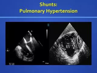
Echocardiography (Ultrasonic Cardiography) This test uses sound waves to track the structure and function of the heart. It can compose images of a beating heart on a monitor and detects: • heart’s thickness, size and function • motion pattern and structure of the four heart valves • → revealing any potential leakage (regurgitation) • thickening of right ventricle, enlarged right heart • reversal of blood through tricuspid valve • extent of lung damage A specific echocardiogram, Doppler ultrasound, is sometimes used to determine pulmonary artery pressure. Exercise echocardiogram – determine how well heart works under stress
Electrocardiogram (ECG) ECG is a record of the electrical activity produced by the heart. Abnormal rhythms (arrhythmias) may indicate that the heart or part of the heart is undergoing unusual stress. Exercise ECG helps evaluation of performance of the heart during exercise, for example, walking on a treadmill in the examination room.
Pulmonary Angiogram Used to measure circulation in the lungs and to visualize clots in the lung on x-rays. The test involves insertion of a thin catheter into the pulmonary artery through which an iodine dye is injected. Image of any blood clots present in the lung can be observed and circulation of blood through lung’s blood vessels can be tracked.
Perfusion Lung Scan Uses small amounts of radioactive tracers (radioisotopes) to study blood flow in the lungs. Radioisotopes are attached to radiopharmaceuticals which are then injected into a vein the arm. A gamma camera takes pictures of blood flow in the lungs’ blood vessels. It is generally used to determine whether blood clots may be causing symptoms of PH. Pulmonary Function Tests Non-invasive tests to measure how much air your lungs can hold and the airflow in and out of your lungs. They can also measure the amount of gases exchanged across the membrane between the lung wall and capillary membrane. During the tests, the patient will be asked to blow into a spirometer. An abnormality here may be amongst the first indication of PH.
Computerized Tomography (CT) Organs can be scanned in two-dimensional ‘slices’. Split-second computer processing creates images as a series of very thin x-ray beams pass through the body. A contrast medium is used to help visualization. The fast CT machine can scan arteries in less than 20 seconds as opposed to 20 minutes for a standard CT. Speed is important because it allows the dye to be visualized while still in the arteries. Magnetic Resonance Imaging (MRI) Uses no x-rays but instead, a computer creates tissue ‘slices’ from data generated by a powerful magnetic field and radio waves. Although not yet routinely used to diagnose PH, it is showing great value in assessing the pulmonary arteries. It cannot, however, measure artery pressure.
Other screening or diagnostic methods • exercise testing • ventilation-perfusion (V/O) scanning • arterial blood gas studies • central hematocrit count • serum glucose and calcium levels count • platelet count • hyperoxia (100% oxygen) challenge test
Until recently, nothing much could be done for people with pulmonary hypertension. Before 1990, there were very few treatments available for PH and the survival rate was approximately two to four years. Since then, a number of exciting new treatments that are able to slow progression of the disease and may even reverse some of the damage to lungs and heart are gradually becoming available. Some people do well on drugs; others may need a transplant. Some patients might also require supplemental oxygen delivered through nasal prongs or a mask if breathing becomes difficult whereas some need oxygen around the clock. In severely affected cases, a heart-lung, single lung of double lung transplantation may be appropriate.
PH patients respond differently to different medicines that are prescribed to dilate or relax blood vessels and no one drug can be said to be consistently effective in all patients. Because individual reactions vary, different drug have to be tried before chronic or long-term treatment begins. During the course of disease, the amount and type of medicine may also have to be changed. To find out which medicine works best for a particular patient, the drugs should be evaluated via cardiac catheterization. This way, they can see the effect of the medicine on the patient’s heart and lungs. They can also adjust the dose to reduce the side effects such as systemic low BP, nausea, angina, headache etc that may occur. To determine whether a drug is improving a patient’s condition, both the pulmonary pressure and the amount of blood being pumped by the heart (cardiac output) must be evaluated.
Calcium Channel Blockers • blocks entry of calcium into muscle cells of heart & arteries • improve ability of heart to pump blood • relaxes smooth muscle in walls of heart and blood vessels • amlodipine (Norvasc), diltiazem (Cardizem, Tiazac), nifedipine (Adalat, Procardia), nicardipine (Cardene) etc. • only small number of people with PH respond to them • side effects – constipation, nausea, headache, rash, edema, drowsiness, dizziness, low blood pressure
Blood Vessel Dilators • Prostacyclin • substance that acts like a hormone (prostaglandin) • imitates behaviour of natural prostacyclin • powerful vasodilator and anti-clotting agent • prevent blood clots from forming • given intravenously through catheter • bridge to help those waiting for transplant • Epoprostenol (Flolan) - 1st vasodilator approved by FDA • Ilopost - inhaled through nebulizer • Treprostinil - injected under skin • side effects - jaw pain, nausea, leg cramps etc • need comprehensive follow-up care
Endothelin Receptor Antagonists • available in pill form • reverse effect of endothelin (blood vessels constriction) • Bosentan (Tracleer) - may improve stamina of people with PH • not for pregnant women • need monthly liver monitoring - risk of liver complications Phosphodiesterase Inhibitors • Revatio – contains sildenafil • same active ingredient used in Viagra • blocks the enzyme phosphodiesterase • accentuates actions of nitric oxide • opens blood vessels in the lungs - dilation • side effect - vision problems
Anticoagulant • warfarin (Coumadin) • prevent formation of blood clots within pulmonary arteries • risk of bleeding complications – prevent normal blood coagulation • periodic blood tests – check how well the drug is working • more than 100 drugs can interact with anticoagulants Diuretics • water pills • eliminate excess fluid from body • reduces amount of work heart has to do • limit fluid buildup in the lungs • improve exchange of gases in lungs
Oxygen • oxygen therapy • especially for those who live in high altitude or have sleep apnea • continuous use of oxygen through nasal prongs/oxygen mask • relieve shortness of breath Cardiotonics • strengthen the contractions of the heart • heart does not need to beat as often to circulate adequate blood for body
Transplants • surgical interventions – considered only in extreme cases • treatment for severe secondary PH if treatment of the underlying • disorder fails • surgically replace damage organs with healthy donated organs • lung and/or heart transplantation • most common : single-lung transplant, fewer complications than double-lung or heart-lung transplant • lung transplant - improvement in structure and functioning of right ventricle • major risks : rejection of transplanted organ, serious infection • take immunosuppressant drugs for life – help reduce chance of rejection • survival rate is about 60% per year and 37% per 5 years
Other treatment procedures • Dilation Atrial Septostomy • experimental procedure • use in patient with severe PH • makes a small hole in the heart, slowly enlarging it to relieve some of the pressure in the heart’s right side • shunts blood across the atrial septum and into the left side of the heart • similar to balloon atrial septostomy – naturally occurring hole present at birth is enlarged to help those with congenital heart defects Other areas of research for treatment of PH includes gene therapy and stem cell research.
Pregnancy and PH The consensus of medical opinion is that PH and pregnancy is very dangerous. The life of the mother and baby are put at great risk. Pregnancy can really take its toll on a woman’s body. For example, heart rate speeds up and the immune system does not work quite as well. For a woman whose body already has to deal with a severe illness, pregnancy can actually have catastrophic consequences. The risk of pregnancy-related death in women with PH is substantial – reported to be as high as 30-50%. Some drugs commonly used to treat PH can be harmful to the developing fetus (e.g. warfarin). Because of this twofold risk to both mother and baby, use of some form of birth control to avoid pregnancy is strongly advised in women of childbearing age with PH.