Download
1 / 35
370 likes | 506 Vues
Explore the detailed boundaries, walls, contents, and structures of the axilla. Learn about nerve supply, injuries, lymphatic drainage, and associated anatomical features in this comprehensive guide.

E N D
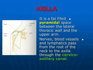
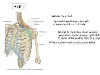
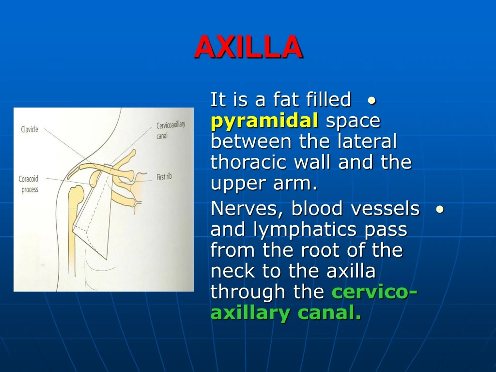
AXILLA • It is a fat filled pyramidal space between the lateral thoracic wall and the upper arm. • Nerves, blood vessels and lymphatics pass from the root of the neck to the axilla through the cervico-axillary canal.
BOUNDARIES • The axilla has : • 1. Apex : it isbounded by: • In front : clavicle. • Behind : upper border of the scapula. • Medially : outer border of the first rib.
BOUNDARIES • 2. Base (lower end) • It is formed by the hairy axillary skin. • Its boundaries are: • In front :anterior axillary fold. • Behind :posterior axillary fold. • Medially:chest wall.
WALLS • The axilla has the following walls : • Anterior. • Posterior. • Medial. • Lateral. • The base is stretched between the anterior and posterior walls.
ANTERIOR WALL • Is formed by : • 1. Pectoralismajor. • 2. Pectoralisminor. • 3. Subclavius & • Clavipectoral fascia.
POSTERIOR WALL • Formed by : • 1. Subscapularis. • 2. Latissmus dorsi. • 3. Teres major.
MEDIAL WALL • It is formed by : • 1.Upper (4) ribs. • 2.Intercostalmuscles. • 3.Serratus anterior.
SERRATUS ANTERIOR • Origin : • Outer surface of the upper (1-8) ribs. • Insertion : • Medial border of the scapula (especially the inferior angle).
SERRATUS ANTERIOR • Nerve supply : • Long thoracic nerve. • Action : • 1.Holds the scapula against the thoracic wall. • 2. protraction of the scapula. • 3. Rotation of thescapula (inferior angle).
WINGED SCAPULA • Injury to the long thoracic nerve (in radical mastectomy) causes paralysis of serratus anterior. • The medial border and inferior angle of the scapula will no longer be kept closely applied to the chest wall.
WINGED SCAPULA • It will protrudeposteriorly. • The patient has difficulty in raising the arm above the head(difficult in rotation of the scapula).
LATERAL WALL • It is narrow. • It is formed by : • 1. Bicipitalgroove of the humerus. • 2. Biceps. • 3. Coracobrachialis.
CONTENTS • 1. Axillary artery. • 2. Axillary vein. • 3. Cords of brachial plexus. • 4. Axillary lymphnodes. • 5. Fat. • 6. Axillary sheath.
AXILLARY ARTERY • Beginning : • At the outer border of the 1st rib as a continuation of thesubclavian artery. • Termination : • Lower border ofteres majorby becoming the brachial artery.
AXILLARY ARTERY • Course : • It is divided bypectoralis minorinto three parts.
RELATIONS • The artery is enclosed throughout its course with the brachial plexus and the axillary vein with a sheath of deepfascia (Axillary sheath).
1ST PART • It extends from the beginning of the artery to the upper border of pectoralis minor. • Anterior :skin, fascia and pectoralis major. • Posterior : long thoracic nerve.
1ST PART • Medial :axillary vein. • Lateral :three cords of the brachial plexus.
2ND PART • It is behind pectoralis minor. • Anterior :skin, fascia, pectoralis minor and major. • Posterior :posterior cord of the brachial plexus, subscapularis.
2ND PART • Medial :medial cord of the B.P. and axillary vein. • Lateral :lateral cord of the B.P.
3RD PART • It begins at the lower border of pectoralisminor and ends at the lower border of teres major by becoming the brachial artery. • Anterior :pectoralis major, medial root of median nerve.
3RD PART • Posterior: • Muscles:subscapularis, teres major & latissmusdorsi. • Nerves:axillary and radial.
3RD PART • Lateral : • Nerves musculocutaneous. • lateral root of median nerve. • Muscles : biceps and coracobrachialis.
3RD PART • Medial:Axillary vein. • Nerves :ulnar and medial cutaneous nerve of the arm.
BRANCHES • First part : superior (highest) thoracic. • Second part : • A. Thoracoacromial. • B. Lateral thoracic.
BRANCHES (3RD PART) • 1.Anterior circumflexhumeral. • 2.Posterior circumflexboth anastomosearound the surgical neck of the humerus). • 3. Subscapular : gives circumflex scapular(share in the anastomosis around the scapula)
AXILLARY VEIN • Formation : • Basilic vein + venae comitantes of the brachial artery. • Beginning : • Lower border of teres major. • Course : • It runs along the medial side of the axillary artery.
AXILLARY VEIN • Course : • It runs along the medial side of the axillary artery. • Trmination : • At outer border of the 1st rib. • It becomes thesubclavian vein.
AXILLARY LYMPH NODES • They are (6) groups. • They drain : • 1. Lateral quadrants of the breast. • 2. Thoracoabdominal walls above umbilicus. • 3. Upper limb.
AXILLARY LYMPH NODES • 1. Pectoral (Anterior) • Lie behind pectoralis major, along lower border of pectoralis minor. • It receives lymph from: • 1.Lateral quadrants of the breast. • 2.Abdominal wall above umbilicus.
AXILLARY LYMPH NODES • 2. Subscapular (Posterior) • Lie in front of subscapularis. • Receives lymph of the back down to the iliac crest. • 3. Lateral : • Lie along medial side of the axillary vein. • Receives from the upper limb except its lateral side.
AXILLARY LYMPH NODES • 4. Central : • Lie in the axillary fat. • Receives from the above three groups. • 5.Infraclavicular (deltopectoral): • Lie in the groove between pectoralis major and deltoid (outside axilla).
AXILLARY LYMPH NODES • 5.Infraclavicular (deltopectoral): • Receives fromlateral sideof: • Arm, forearm and hand.
AXILLARY LYMPH NODES • 6. Apical : • Lie at the apex of the axilla , at the lateral border of the 1st rib. • It receives efferent vessels from all the other lymph nodes. • They drain into the Subclavianlymph trunk.
DRAINAGE • On right side : • The trunk drains into rightlymph trunk. • On left side : • It drains into thoracic duct.