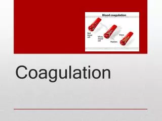
Coagulation Mechanisms
Coagulation Mechanisms. TEXTBOOK OF MEDICAL PHYSIOLOGY GUYTON & HALL 11 TH EDITION UNIT VI CHAPTER 3 6 Dr Mohammed Alotaibi MRes , PhD (Liverpool, England) Assist . Professor Department of Physiology College of Medicine King Saud University . Objectives.

Coagulation Mechanisms
E N D
Presentation Transcript
Coagulation Mechanisms TEXTBOOK OF MEDICAL PHYSIOLOGY GUYTON & HALL 11TH EDITION UNIT VI CHAPTER 36 Dr Mohammed Alotaibi MRes, PhD (Liverpool, England) Assist. Professor Department of Physiology College of Medicine King Saud University
Objectives At the end of this lecture student should be able to: • Recognize the different clotting factors • Understand the role of calcium ions during clotting cascades. • Describe the cascades of intrinsic and extrinsic pathways for clotting. • Recognize process of fibrinolysis and function of plasmin • Recognize some conditions causing excessive bleeding • Understand some important anticoagulants and their mechanism of action
Mechanism of Blood Coagulation • A crucial physiological balance exists between factors promoting coagulation (procoagulants) and factors inhibiting coagulation (anticoagulants). • Coagulation of blood depends on the balance between these two factors. • Disturbances in this balancecould lead to thrombosis or bleeding
Hemostasis: • prevention or stoppage of blood loss. • Hemostatic Mechanisms: • Vessel wall (Vasoconstriction) • Platelets (Production and activation, Platelets Plug formation) • Blood coagulation • Clot formation (intrinsic & extrinsic pathways) • 4. Fibrinolysis
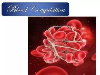
Coagulation: Formation of fibrin meshwork (Threads) to form a CLOT
Activated Factor X Prothrombin activator Prothrombin Thrombin Fibrinogen Fibrin
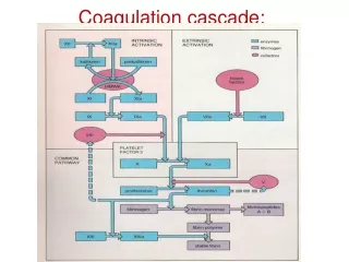
The Coagulation Cascades Intrinsic pathway Extrinsic pathway Activators: Collagen and damaged endothelium Common pathway Activator: Tissue factor (III) (Thromboplastin) V, Ca, Phospholipids XX activated FibrinogenFibrin monomer XII XII activated VII VII activated XI XI activated IX IX activated Prothrombin Thrombin III, Ca, Phospholipids Prothrombin activator Ca VIII, Ca, Phospholipids 3-6 minutes 12-16 seconds XIII Ca Fibrin fibers polymers (Insoluble)
Thrombin • Thrombin changes fibrinogen to fibrin • Thrombin is essential in platelet morphological changes to form primary plug • Thrombin stimulates platelets to release ADP & thromboxane A2; both stimulate further platelets aggregation • Activates factor V, VIII, XIII
Blood coagulation (clot formation) • A series of biochemical reactions leading to the formation of a blood clot within few seconds after injury • Prothrombin (inactive thrombin) is activated by a long intrinsic or short extrinsic pathways • This reaction leads to the activation of thrombin enzyme from inactive form prothrombin • Thrombin will change fibrinogen (plasma protein) into fibrin (insoluble protein)
Intrinsic pathway • The trigger is the activation of factor XII by contact with foreign surface, injured blood vessel, and glass. • Activated factor XII will activate factor XI • Activated factor Xl will activate IX • Activated factor IX + factor VIII + platelet phospholipid factor (PF3)+ Ca activatefactor X • Following this step the pathway is common for both intrinsic and extrinsic
Extrinsic pathway • Triggered by material released from damaged tissues (tissue thromboplastin) • Tissue thromboplastin + VII + Ca activate X Common pathway • Activated factor X + factor V +PF3 + Ca activateprothrombin activator; aproteolytic enzyme which activates prothrombin. • Activated prothrombinactivatesthrombin • Thrombin acts on fibrinogen and change it into insoluble thread like fibrin. • Factor XIII + Ca strong fibrin (strong clot)
Activation of Blood Coagulation • Intrinsic Pathway: all clotting factors present in the blood • Extrinsic Pathway: triggered by tissue factor (thromboplastin) Common Pathway
Fibrinolysis • Formed blood clot can either become fibrous or dissolved. • Fibrinolysis (dissolving) = Break down of fibrin by naturally occurring enzyme plasmin therefore prevent intravascular blocking. • There is a balance between clotting and fibrinolysis • Excess clotting blocking of Blood Vessels • Excess fibrinolysis tendency for bleeding
Fibrinolysis Tissue Plasminogen Activator (t-PA) Thrombin Plasminogen Plasmin Fibrinogen Fibrin FDP* Streptokinase Released from healed tissues and vascular endothelium Urokinase (Protein in the blood) Fibrinolysis FDP*: Fibrin Degradation Products
Plasmin • Plasminis present in the blood in an inactive form plasminogen • Plasmin is activated by tissueplasminogen activators (t-PA) in blood. • Plasmindigests intra & extra vascular deposit of Fibrinfibrin degradation products (FDP) • Unwanted effect of plasmin is the digestion of clotting factors • (α2-antiplasminorplasmin inhibitor) is responsible for inactivating plasmin
Prevention of blood clotting in the normal vascular system • Endothelial surface factors • Smoothness of the ECs. • Glycocalyx layer • Thrombomodulin protein • Fibrin fibers, adsorbs ~ 90% of thrombin to removes it from circulating blood • Antithrombin III, combines the remaining thrombin and removes it from blood • Heparin, combines with AntithrombinIII and quickly removes thrombin from blood - Liver, lungs, mast cells, basophils
Anticoagulants • Anticoagulants for clinical use: • Heparin • - Commercial, extracted from animals • Coumarins • - Warfarin, competitive with vitamin K • - decrease Factors II, VII, IX, X • Prevention of blood coagulation outside the body: • (decrease calcium ion concentration) • Oxalate (precipitation, toxic ) • Citrate (deionizer) • EDTA (Chelating agent)
Conditions that cause excessive bleeding • Vitamin K Deficiency • Factor II, Factor VII, Factor IX, Factor Xrequire vitamin K for their synthesis • Hepatitis, Cirrhosis, acute yellow atrophy AND GI disease • Hemophilia • ↑ bleeding tendency. • Affects males. • 85% due to Factor VIII deficiency (hemophilia A),and 15% due to Factor IX deficiency (hemophilia B). • Thrombocytopenia • Very low number of platelets in blood (< 50,000/μl) • Thrombocytopenia purpura, hemorrhages throughout all the body tissues • IdiopathicThrombocytopenia, unknown cause.
The END Thrombocytopenia purpura