Download

1 / 22
220 likes | 407 Vues
Articular System (Joints) ANHB 2212 – 2007. Avinash Bharadwaj. Joints. “Articulation” Articulatio / Arthron Arthrology Where two or more bones or parts of bones unite… How they are joined together… the tissues that unite bones. Joint Classification. Structural

E N D
Articular System (Joints)ANHB 2212 – 2007 Avinash Bharadwaj
Joints • “Articulation” • Articulatio / Arthron Arthrology • Where two or more bones or parts of bones unite… • How they are joined together…the tissues that unite bones
Joint Classification • Structural • The tissues that unite bones • Functional • Mobility or otherwise • Synovial joints – axes of movements • No perfect system (Explained as we go!) • Arbitrary and confusing terms • Overlapping terminology • Latin terminology and anglicised versions • The choice is yours!
Tissues Uniting Bones • Fibrous tissue • Unossified tissue between membrane bones (Explained fully next week!) • Ligaments (Dense connective tissue!) • Cartilage • Hyaline cartilage • White fibrocartilage • Fibrous capsule with a cavity • Special features for lubrication, movement and stability
Movable Joints • “Movability” (mobility…?) is a relative term! • Movement is always associated with restrictions • Active movements – by muscles • Movements due to other factors • Shapes of bony surfaces • Tensions in ligaments • Other external factors (pelvic joints in childbirth) • Passive movements in clinical examination
Fibrous Joints • Sutures (“stitches”) • Between flat bones of the skull Surface view Section (Magnified!) Periosteum Bone
R U Fibrous Joints • Syndesmoses • Inferior tibiofibular joint (Not shown here) • Interosseous membranes • Separation of compartments • Additional areas for muscle attachments • Some other ligaments… • Gomphoses • Tooth and bone • “Peg-and-socket”
Cartilaginous Joints • Synchondrosis – Hyaline cartilage • Growth mechanism synostosis • Between primary and secondary centres • Between named bones (e.g. sphenoid + occipital) (Week 12 Histology!) • Symphysis – White fibrocartilage • WFC is deformable • Partially movable joints • Occasional cavities – ? Evolution of synovial joints Primary and secondary cartilaginous joints : terms best avoided. Functionally two distinct entities.
Synovial Joints • “Freely movable”…? • Freedom is relative! • More movable than any other type…! • Fibrous tissue bond, with a cavity
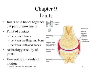
Synovial Joints – Basic Features • The bones – articular surfaces • Smooth… but not quite! • Articular cartilage • Fibrous capsule • Ligaments • Capsular thickening • External • Synovial membrane
Synovial Joints – Articular Cartilage • Hyaline (WFC in membrane bones) • No perichondrium • Variable thickness • Depends on the type of joint • In curved surfaces • Convex – thick in the centre • Concave – thin in the centre
Synovial Joints – Capsule Etc • Capsular attachment • Thickening of capsule : “Intrinsic” ligaments • Areas of strength / weakness • Capsule may be loose in places • Other ligaments (“extracapsular”) • Synovial membrane and fluid
Intra-articular discs • White Fibrocartilage • Reduce incongruity • Modify movements • Spread of synovial fluid – reduce “drag” • Incomplete discs / menisci
Other Intra-articular Structures • Ligaments • Cruciate ligaments – knee joint • Tendons • Long head of biceps – shoulder joint • Popliteus – knee joint • Fat pads • Such structures are covered by synovial membrane.
Synovial Joints – Subtypes • Functional • Degrees of freedom • Uniaxial, biaxial, multiaxial • Structural • Shapes of bony surfaces
Synovial Joints – Subtypes • Plane : Sliding movements(“Non-axial joint”) • Hinge : Uniaxial (e.g. elbow) • Ball-and-socket : Multiaxial (Shoulder, hip) • Saddle : Two planes of movement + combinations • Bicondylar : Two pairs of surfaces • Pivot : Osseofibrous ring + rotating bone Nothing is perfect – do not expect geometrical shapes!
Two Types Illustrated Pivot joint – Osseofibrous ring and head of radius. Bicondylar joint – Knee Two pairs of articular surfaces
Bursae • Muscles / tendons in close proximity – friction • Bursa (Latin for purse!) • Thin walled connective tissue bag • Lining of synovial membrane • Between joint capsule and tendon / muscle, bone and tendon / muscle, or between muscles / tendons. • Bursitis – painful • Some bursae communicate with joint cavity
Joint Stability • Movement and stability – a compromise • Factors in stability • Bony configuration • Capsule and ligaments • Tendons and muscles • Sometimes blend with capsule – rotator cuff
Nerves • Rich sensory innervation • Capsule, ligaments, periosteum • Same nerves as those that supply muscles • Joint pain • Bony injuries • Capsular / ligamentous injuries • Synovial membrane • And more…
Blood Vessels • Rich blood supply • All parts except cartilages • Articular cartilage • By diffusion through synovial fluid • Blood vessels of surrounding areas (capsule, bone) Last Slide