Download
1 / 104
1.05k likes | 1.83k Vues
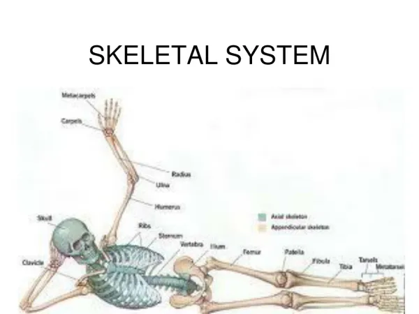
SKELETAL SYSTEM. HUMAN SKELETAL SYSTEM. CLASSIFICATION OF BONES There are 206 bones in the human body The human skeleton is divided into the Axial and Appendicular skeletons Skeletal system also includes: Joints Cartilages Ligaments. The Functions of Bones:. Support

E N D
HUMAN SKELETAL SYSTEM CLASSIFICATION OF BONES • There are 206bones in the human body • The human skeleton is divided into the Axialand Appendicular skeletons • Skeletal system also includes: • Joints • Cartilages • Ligaments
The Functions of Bones: • Support • Protection of ORGANS • Movement– bones attached to muscles • Mineral Storage such as CALCIUM & PHOSPHORUS • PRODUCE RED BLOOD CELLS • STORE FAT
Classification of Bones • Two main types of bone • Compact = dense, looks smooth • Very hard • Spongy = made of small needlelike bone pieces, lots of open space • Slightly soft • Bones are also classified by shape
Classification of Bones by Shape • LONG • Bones of limbs except wrists, ankles, and kneecap • Mostly compact bone • Short • Bones of wrist and ankles, kneecap • Cube-shaped, mostly spongy bone • Flat • Bones of skull, ribs, and sternum • Flat, two layers of compact sandwiching spongy • IRREGULAR • vertebrae
Long Bone Structure Bones are organs because they contain several types of tissue including: • CONNECTIVE, NERVOUS, muscle and epithelial
The structure of a typical long bone includes: 1. Compact Bone=the EXTERNAL layer of the bone MICROSCOPIC VIEW OF COMPACT BONE MACROSCOPIC VIEW OF COMPACT BONE
LONG BONE STRUCTURE 2. Spongy Bone =internal bone structure that is POROUSand contains red and YELLOWbone marrow SPONGY BONE SPONGY BONE
LONG BONE STRUCTURE • 3. Diaphysis= the SHAFTor long axis of the bone composed of compact bone • Diaphysis is covered by the periosteum • Periosteum = connective tissue membrane DIAPHYSIS
LONG BONE STRUCTURE 4. Medullary Cavity= or marrow cavity, in adults it contains FAT(yellow marrow) and is found inside the DIAPHYSIS
LONG BONE STRUCTURE 5. Epiphyses = the ENDS of the bones • Thin layer of compact bone enclosing an area filled with spongy bone EPIPHYSES EPIPHYSES
LONG BONE STRUCTURE 6. ARTICULAR CARTILAGE = cushions the opposing bone ends during joint movement and absorbs stress • Instead of periosteum
LONG BONE STRUCTURE 7. Epiphyseal Line = thin line of bone between epiphyses and diaphysis that looks different • Remnant of the epiphyseal plate, a disc of cartilage that grows during childhood to LENGTHEN the bone
Label the Parts of a Long Bone Spongy bone, diaphysis, compact bone, epiphyses, articular cartilage, periosteum, epiphyseal line, medullary cavity Epiphyses Epiphyses Diaphysis Medullary cavity Spongy bone Epiphyseal line Articular cartilage Articular cartilage Compact bone Periosteum
Short, Irregular and Flat Bone Structure • The structure of the short, irregular and flat bones is the sameas the long bones in the following ways: • Both contain periosteum-coveredCOMPACTbone on the outside • Both contain endosteum-coveredSPONGYbone on the inside 3. Both contain bone MARROW
Short, Irregular and Flat Bone Structure • The structure of the short, irregular and flat bones is different from the long bones in the following ways: 1. No DIAPHYSIS(shaft) 2. No EPIPHYSES (bone ends) 3. The internal layer of spongy bone is called DIPLOE
Irregular Bone Structure COMPACT BONE SPONGY BONE = DIPLOE
Bone Markings • Bones usually have what look like to be bumps, holes and ridges • Actually bone markings • Where muscles, tendons, and ligaments were attached, and where blood vessels and nerves passed through • Two categories of bone markings: • Projections/processes = grow out from bone • Depression/cavities = indentations • Table 5.1 on pg. 140 in your book has a list of these
Compact Bone • Even though it is more dense than spongy bone, there are still openings and passageways throughout compact bone • Nerves and blood vessels travel through these passageways • Provides living bone cells with nutrients
Bone Cells • Osteocyte = mature bone cell, found in lacunae or cavities • Lacunae are arranged in concentric circles called lamellae • Lamellae surround the central (Haversian) canals • Osteon = central canal + matrix rings
Central Canals • Haversian canals run lengthwise through the bone, carrying blood vessels and nerves to rest of bone • Canalicuili = tiny canals coming out of the central canals to reach all lacunae • Forms a transport system that connects all bone cells to nutrient supply • Perforating (Volkman’s) canals = travel perpendicular to the shaft to connect central canal to the outside of bone
Ossification • The embryonic skeleton is made of cartilage • Over time the cartilage turns into bone • Ossification = process of bone formation • Two steps to ossification: • Osteoblasts cover hyaline cartilage with bone matrix • Hyaline cartilage is digested, which opens up the medullary cavity
Ossification Continues Throughout Childhood • By birth, the entire skeleton should be converted to bone • Except the articular cartilage and epiphyseal plates • These areas continue to form new cartilage throughout your growing process • Overtime, the cartilage is replaced with bone
Bones Also Grow Wider • Bones don’t just grow longer, they get wider too • Osteoblasts add bone tissue to external surface of diaphysis wall • Appositional growth = increasing diameter of bones • Bone growth is controlled by hormones • Growth hormone, sex hormones
Bone Remodeling • Bones are constantly being remodeled • Bones remodel in response to two things: • Calcium levels in the blood • If too little calcium, osteoclasts break down bone and put it into blood • If too much calcium, its deposited in the bone • Pull of gravity and muscles on skeleton • The more you exercise, the bigger/stronger your bones are
Fractures • Fracture = break in the bone • There are many different types of fractures • Fractures are classified by four things • Position after fracture • Completeness of break • Orientation of break • Penetration through skin
1. Position of Bone After Fracture • Nondisplaced fractures= the bones retain their normal position • Displaced fractures = the bone ends are out of normal alignment
2. Completeness of Break • Incomplete fracture = if the bone is NOT completely broken all the way through • Complete fracture =if the bone is completely broken through
INCOMPLETE & COMPLETE FRACTURES Incomplete Fractures Complete Fractures
3. Orientation of Break to Long Axis of Bone • Linear fracture =the break is parallel to the long axis of the bone • Transverse fracture =the break is perpendicular to the bone’s long axis • Oblique fracture = the break is diagonal to the long axis of the bone
LINEAR, TRANSVERSE, & OBLIQUE FRACTURES Linear Fracture Transverse Fracture Oblique Fracture
4. Penetration of Skin • Open fracture (compound) = the bone ends penetrate the skin • Closed fracture (simple) = the bone ends do NOT penetrate the skin
Common Types of Fractures Fracture Type Description Found where/ or in who? 1.Comminuted 2.Spiral 3.Depressed 4.Compression 5.Epiphyseal 6.Greenstick BONE FRAGMENTS ELDERLY TWISTING ATHLETES BROKEN BONE PRESSED IN SKULL CRUSHED VERTEBRA SEPARATION AT EPIPHSEAL LINE EPIPHYSEAL LINE INCOMPLETE CHILDREN
Fracture Repair • Fracture is treated by reduction • Reduction = realignment of broken bone ends • Two types: • Closed Reduction = the bone ends are coaxed back into position by the physician’s hands • Open Reduction = the bone ends are secured together surgically with pins or wires • Healing time is usually 6-8 weeks
Four Steps to Fracture Repair • Hematoma formation • Fibrocartilage callus formation • Bony callus formation • Bone remodeling
1. Hematoma Formation • Blood vessels are ruptured when bones break • Creates a hematoma • Hematoma = blood-filled swelling • Lack of nutrients to surrounding bone cells causes them to die
2. Fibrocartilage Callus Formation • Phagocytes (WBCs) “clean out” the injured area and dispose of dead tissue • Fibroblasts and osteoblasts migrate to the fracture from the periosteum & endosteum • Fibroblasts secrete collagen fibers to heal the fracture and osteoblasts construct new bone • Creates the fibrocartilage callus, which acts as a “splint”
3. Bony Callus Formation • More osteoblasts migrate to the area • Osteoblasts gradually replace fibrocartilage callus with new spongy bone • Creating a bony callus
4. Bone Remodeling • Bony callus is remodeled in response to mechanical stresses put on the bone • Osteoclasts remodel the bony callus by removing excess bone • Osteoblasts create compact bone • This phase lasts for several months to years following the injury


