Download

1 / 86
930 likes | 1.53k Vues
IABP- Instrumentation , Indications and Complications. Dr Sajeer KT Senior Resident Dept.of Cardiology, MCH Calicut. Intra aortic balloon counter pulsation( IABP):. Temporary support for the left ventricle by mechanically displacing blood within the aorta.

E N D
IABP- Instrumentation, Indications and Complications Dr Sajeer KT Senior Resident Dept.of Cardiology, MCH Calicut
Intra aortic balloon counter pulsation( IABP): Temporary support for the left ventricle by mechanically displacing blood within the aorta Most common and widely available methods of mechanical circulatory support Concepts: - Systolic unloading - Diastolic augmentation Traditionally used in surgical and non surgical patients with cardiogenic shock
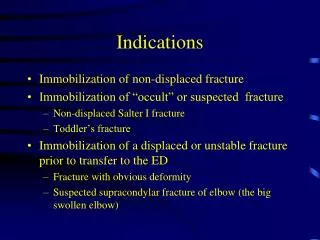
Indications for IABP 1. Cardiogenic shock: - Associated with acute MI - Mechanical complications of MI - MR , VSD 2. In association with CABG : Preoperative insertion - Patients with severe LV dysfunction - Patients with intractable ischemic arrhythmias Postoperative insertion - Postcardiotomy cardiogenic shock 3. In association with nonsurgical revascularization: • Hemodynamically unstable infarct patients • High risk coronary interventions • - severe LV dysfunction, LMCA, complex coronary artery disease 4. Stabilization of cardiac transplant recipient before insertion of VAD Post infarction angina Ventricular arrhythmias relathed to ischemia
Contraindications to IABP • Severe aortic insufficiency • Aortic aneurysm • Aortic dissection • Limb ischemia • Thrombo embolism
Cardiac cycle LV contraction: - Isovol. Contraction (b) - maximal ejection (c) LV relaxation: - start of relaxation and reduced ejection (d) - isovol.relaxation (e) LV filling: - LV filling , rapid phase (f) - slow LV filling (g) - atrial systole( a)
The IAB Counter pulsation system - two principal parts • A flexible catheter -2 lumen • first - for distal aspiration/flushing or pressuremonitoring • second - forthe periodic delivery and removal of helium gas to a • closed balloon. • A mobile console • system for helium transfer • computer for control of the inflation and deflation cycle
HEMODYNAMIC EFFECTS — Inflation and deflation of the balloon • Blood is displaced to the proximal aorta by inflation during • diastole. • Aortic volume ( afterload) is reduced during systole through a vacuum effect created by rapid balloon deflation
Expected changes with IABP support in hemodynamic profile in patients with Cardiogenic shock • - Decrease in SBP by 20 % • Increase in aortic Diastolic Press. by 30 % ( raise coronary blood flow) • Increase in MAP • Reduction of the HR by 20% • Decrease in the mean PCWP by 20 % • - Elevation in the COP by 20%
IABP catheter: • 10-20 cm long polyurethane bladder • 25cc to 50cc capacity • Optimal 85% of aorta occluded (not 100%) • The shaft of the balloon catheter contains 2 lumens: • - one allows for gas exchange from console to • balloon • - second lumen • - for catheter delivery over a guide wire • - for monitoring of central aortic pressure • after installation.
Benefits of larger volume IABs More blood volume displacement More diastolic augmentation More systolic unloading
IABP Kit Contents Introducer needle • Guide wire • Vessel dilators • Sheath • IABP (34 or 40cc) • Gas tubing • 60-mL syringe • Three-way stopcock
STEP BY STEP- IABP insertion • Connect ECG • Set up pressure lines • Femoral access – followed by insertion of the supplied • sheath(7.5 F) • 0.030 inch supplied J-shaped guide wire to the level of the • aortic arch (LAO view)
Take the entire catheter and T handle as one unit (DO NOT disconnect one-way valve when removing the extracorporeal tubing from the tray.) Pull out the T- handle only as shown
Inserting the Balloon catheter • • Remove stylet/aspirate/Flush • • Insert the balloon only over the guide wire • • Hold the catheter close to skin insertion point • • Advance in small steps of 1 to 2 cm at a time and • stop if any resistance. • • The IABP should advance freely • Many vascular complications occur during insertion itself • Resistance during insertion either indicates PVOD, or dissection • - Kinking of IABP » improper inflation/deflation
Positioning • The end of the balloon should be just distal (1-2 cm) to the takeoff of the • left subclavian artery • - Position should be confirmed by fluoroscopy or chest x-ray
Connecting to console: • - Connect helium gas tube to the console via a long extender • Open helium tank. • The central lumen of the catheter is flushed and connected to pressure • tubing with 3 way and then to a pressure transducer to allow for • monitoring of central aortic pressure. • - Zero the transducer Initial set-up: - Once connected properly the console would show ECG and pressure waveforms. - Check Basal mean pressure - Make sure the setting is at “auto” - Usually IABP started at 1:1 or 1:2 augmentation - Usually Augmentation is kept at maxim
Trigger modes Trigger : - Event the pump uses to identify the onset of cardiac cycle (systole) - Pump must have consistent trigger in order to provide patient assist - If selected trigger not detected, counter pulsation will interrupted 1.ECG - uses the slope of QR segment to detect triggering point 2. AP(Arterial pressure wave) - Systolic upstroke of the arterial pressure wave form is the trigger 3. IN(Internal trigger)
ECG signal – most common • Inflation - middle of T wave • Deflation – peak of R wave • Pacer (v/a) • Arterial waveform • An intrinsic pump rate (VF, CPB)
Initial settings Auto Operation Mode Automatic lead and trigger selection Automatic and continuous inflation and deflation timing management - User has ability to fine-tune deflation timing Automatic management of irregular rhythms Semi-Auto Operation Mode Operator selects most appropriate lead and trigger source
The “normal” augmented waveform Increased coronary perfusion
Not all Sub optimal augmentation is due to Timing errors/kinks
Factors affecting diastolic augmentation Patient - Heart rate - Mean arterial pressure - Stroke volume - Systemic vascular resistance Intra aortic balloon catheter - IAB in sheath - IAB not unfolded - IAB position - Kink in the IAB catheter - IAB leak - Low helium concentration Intra aortic balloon pump - Timing - Position of IAB augmentation control
How to check waveform is acceptable ? • First change from 1:1 to 1:2 augmentation
How to check waveform is acceptable ? • First change from 1:1 to 1:2 augmentation • Check the dicrotic notch • See if augmentation starts at that point • This should produce a sharp “V” at inflation.
How to check waveform is acceptable ? • First change from 1:1 to 1:2 augmentation • Check the dicrotic notch • See if augmentation starts at that point • This should produce a sharp “V” at inflation. • Check if diastolic augmented wave is › systolic wave
How to check waveform is acceptable ? • First change from 1:1 to 1:2 augmentation • Check the dicrotic notch • See if augmentation starts at that point • This should produce a sharp “V” at inflation. • Check if diastolic augmented wave is › systolic wave • Confirm if end diastolic wave • following the augmented wave • is less than an non augmented • wave. • Is Deflation slope ok
Late Inflation • Inflation of the IAB markedly after closure of the aortic valve. • Waveform Characteristics: • • Inflation of IAB after the dicrotic notch. • • Absence of sharp V. • Sub optimal diastolic augmentation