Download
1 / 65
750 likes | 1.13k Vues
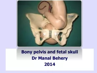
Bony pelvis and fetal skull Dr Manal Behery 2014. Objects. Fetal head Pelvic anatomy Pelvic shapes. Passenger. Fetal head. Landmarks Sutures Fontanelles Diameters. Fetal head. From an obstetrical point of view it’s the most important part: largest

E N D
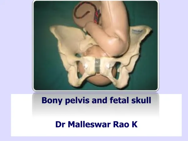
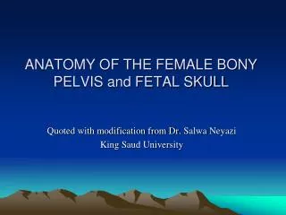
Bony pelvis and fetal skull Dr ManalBehery 2014
Objects • Fetal head • Pelvic anatomy • Pelvic shapes
Fetal head • Landmarks • Sutures • Fontanelles • Diameters
Fetal head From an obstetrical point of view it’s the most important part: • largest • least compressible part of the fetus. • most frequent presenting part
Landmarks • Head is divided into 3areas • (1) the sinciput or brow portion; • (2) the vertex, or top of the head between the 2 fontanelles; • (3) Base large, ossified, firmly united, and noncompressible
Sutures • Membrane-occupied spaces between the cranial bones 1-Sagittal suture: - lies btw the parietal bones -extends in an AP direction btw the fontanelles -divides the head into right and left sides
2-lambdoid suture: • extends from the posterior fontanelle laterally • separate the occipital from the parietal bones.
3-coronal suture: • extends from the anterior fontanelle laterally • separate the parietal and frontal bones.
4- frontal suture: • lies between the frontal bones • extends from the anterior fontanelle to the glabella (the prominence between the eyebrows).
Clinical importance of sutures • Position of fontanelle & sagittal suture can identify attitude and position of vertex. • By plapating the sagittal suture during labour, degree of internal rotation & molding of the head can be noticed. • In deep transverse arrest, this sagittal suture lies transversely at the level of the ischial spines.
Moulding… Reshaping of the fetal skull: Obliteration of the sutures. Overlapping of the bones of the vault: One parietal bone overlaps the other. Both overlap the occipital bone. It accounts for diminution of the biparietal diameter and suboccipitobregmatic diameters by 0.5-1 cm. 0r even more.
A: Well flexed Head • B: Partially Flexed Head • C: Deflexed Head • D: Face Presentation • E: Brow presentation
The anterior fontanelle (bregma) : • diamond shaped area(2 × 3 cm) of unossified membrane formed by the junction of 4 suture.
The posterior fontanelle: • It is the triangular depressed area at the junction of 3 suture: • closes at 6 to 8 weeks of life • Y- or T-shaped
ROT LOT
LOA ROA
LOP ROP LOP
presenting AP diameter in a brow presentation • longest AP diameter of the head
-4. Submentobregmatic (9.5 cm):presenting AP diameter in face presentations
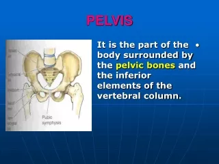
False Pelvis bordered by: • lumbar vertebrae posteriorly • iliac fossa bilaterally • abdominal wall anteriorly. • supports the abdominal contents • after 1st trimester helps support the gravid uterus.
SP Ischial Spine Ischial Tuberosity
The Planes of the pelvis • Plane of the pelvic inlet. • Plane of the cavity: Plane of greatest Pelvic Dimensions • Plane of the mid pelvis (plane of obstetric outlet) • Plane of the Anatomical outlet
(Inlet (Pelvic brim) Bounded by passing with the boundaries of pelvic brim and making an angle of 55o with the horizon (angle of pelvic inclination).
Pelvic inclination:The plane of the pelvic inlet makes an angle of 55 degree with the horizon in the standing position"
The consequences of walking upright… • When a women stands erect: • The pelvic inlet makes an angle of about 55° with the horizon. • The pelvic outlet makes an angle of 15° with the horizon • If the angle made by the inlet is greater than 55° this may make the descent of the fetal head in the pelvis difficult.
The True Conjugate = 11 cm The Obstet. Conjugate = 10.5cm The Diagonal Conjugate = 12 cm
(4) External conjugate: From: The upper anterior margin of the symphysis pubis. To: The depression below the tip of the 5th lumbar spine (20 cm).
(5) Anatomical transverse diameter: Between the farthest points on iliopectineal lines (= 13 cm). It lies 4 cm infornt of the sacral promontory, 7 cm behind the symphysis pubis. (6) Obstetric transverse diameter: It bisects the true conjugate (12.5 cm) TC ATD OTD
(7) The oblique diameters: (12 cm) The right extends from the right joint to the left eminence and vice versa. (8) Sacrocotyloid diameter: (9-9.5 cm) The right diameter ends in the right eminence & vice versa.
The pelvic Cavity • Boundaries: • Above: pelvic brim. • Below: plane of least pelvic dimensions. • Anteriorly: syrnphysis pubis. • Posteriorly: sacrum.
2-pelvic cavity The plane of greatest diameter: bordered by: • the posterior midpoint of the pubis anteriorly • the upper part of the obturator foramina laterally • the junction of the 2nd and 3rd sacral vertebrae posteriorly. The fetal head rotates to the anterior position in this plane
The pelvic Outlet • Anatomical outlet: Lozenge -shaped, bounded by: • Lower border of symphysis pubis. • Pubic arch. • Ischialtuberosities • Sacrotuberous & sacrospinous ligaments. • Tip of the coccyx.
Anterior Sagittal Plane Posterior Sagittal Plane The Plane of the Outlet