Download
1 / 49
490 likes | 748 Vues
Medical Treatment of Retinopathy of type 2 diabetes. José Cunha-Vaz. Coimbra * Portugal. Trends in Diabetes Prevalence. The prevalence of diabetes among US adults increased from 4.9% in 1990 to 7.3% in 2000, an increase of 49%.

E N D
Medical Treatment of Retinopathy of type 2 diabetes José Cunha-Vaz Coimbra * Portugal
Trends in Diabetes Prevalence • The prevalence of diabetes among US adults increased from 4.9% in 1990 to 7.3% in 2000, an increase of 49%. • The worldwide prevalence of diabetes will more than double between 2000 and 2030.
Highest Increase in Diabetes Prevalence Occurs in Younger Adults
Diabetes is associated with multiple vascular complications Hyperglycemia Hypertension Dyslipidemia Obesity Cerebral Ocular • Stroke • Death • Diabetic retinopathy • Cataract • Macular edema • Blindness Atherosclerosis Thrombosis Endothelial dysfunction Hypertension Inflammation Coronary • Myocardial infarction • Heart failure • Death Renal • Nephropathy • End-stage renal disease • Death Peripheral • Claudication • Amputation
Diabetic retinopathy is a common complication of diabetes • Each year, ~20.000 individuals with diabetes become blind in the United States
Limitations of present classifications 1. Do not take into consideration different rates of progression2. Do not include macular edema3. Do not characterize well mild to moderate NPDR- stages that are reversible 4. Assume that all patients progress to Proliferative Retinopathy- Proliferative Retinopathy - Just a response to ischemia? - Independent of diabetes ?
Evolution of Diabetic RetinopathyGeneral Clinical Impression Different evolution in different patients with similar metabolic control and duration of diseaseNot all patients develop persistent macular edema Not all patients develop neovascularization
Treatment to be effective: - Reversible stages of retinal damage- Before vision loss 20 - 35 - 43 ETDRS gradings Need to focus on initial stages D.R. Diabetic Retinopathy
Microaneurysms / Hemorr. RED DOTS - Fundus Photography- Alteration BRB LEAKAGE - F. A. (Qualitative) - Vitreous Fluorometry - Retinal Leakage Analyzer- Alt. hemodynamics BLOOD FLOW - Doppler Flowmeter - Capillary closure FAZ - F.A. - Neuroglial edema THICKNESS - OCT, RTA - Vision changes - VF; ETDRS scale Initial Clinical Alterations – D.R.
Evaluation of Initial Clinical Alterations - Red dots counting - Fundus Photography - Leakage - Vitreous Fluorometry - Retinal Leakage Analyzer - Blood Flow - Flowmeter - FAZ Alterations - Fluorescein Angiography - Retinal Thickness - Retinal Thickness Analyzer OCT
Red dot ID by location Rate of red dot formation and disappearance
Selection Visit 6 Month Visit VS, V6 V6 Perspective Registration (TS) Perspective Registration (T6) VS, V6 VS, V6 V6 VS Reference Fundus
RED DOT COUNTING – Digitized fundus images - Red dots counting – considering specific location - Red dots formation and disappearance rates Index of Progression of Retinal Vascular Damage Number of accumulated red dots years of follow-up Red dot formation rate =
RED DOT COUNTING – Non-invasiveDifferent individuals with same RD grading show Different rates of progression of retinal vascular damage identified by different Red Dot Formation Rates. - Torrent-Solans et al. ARVO 2004 -
LEAKAGE -Measurement of Blood-Retinal Barrier Vitreous Fluorometry Determination of fluorescein penetration into the vitreous across BRB – Flurotron Master Retinal Leakage Analyzer Mapping fluorescein penetration into the vitreous across BRB based on a confocal scanning laser ophthalmoscope
Retinal Thickness RTA II Retinal Thickness Analyzer OCT 3 Optical Coherence Tomography Values expressed as % increases in thickness over mean + 2SD over healthy controls
10-7cm/s Multimodal Macula Mapping
Multimodal Macula Mapping A combination of: - Digital fundus photography – RED DOTS - Fluorescein angiography (FA) – FAZ - Retinal Leakage Analysis (RLA) – LEAKAGE - Capillary Blood Flow (HRF) – BLOOD FLOW - Retinal Thickness Analysis (RTA) – THICKNESS
Multimodal Macula Mapping Morphology Visual function FAZ BRB function/central leaking sites Edema Blood Flow
Follow-up Studies • Initial stages – ETDRS – 20, 35 (stabilized metabolic control) • 3 year follow-up – 14 patients • (Arch. Ophthalmol., 2004) • 2. 6 year follow-up – 57 patients • (IOVS, 2005) • 2 year (6 month intervals) + 4 years (1/year)
Stabilised Metabolic Control First two years of follow-up HgA1C levels and BP medication
D.R. Phenotypes –type 2 Diabetes Phenotype A Slow Progression Minimal Leakage Slow Remodelling Phenotype B Leaky Hemodynamic High Leakage High Flow Rapid progression Phenotype C Ishemic Thrombotic FAZ – Capillary closure Moderate Leakage Decreased Flow Rapid Progression
57 eyes from patients with mild NPDR 35 – Pattern A (61%) – one eye CSME (3%) 12 – Pattern B (21%) – eight eyes CSME (67%) 10 – Pattern C (18%) – six eyes CSME (60%) 6 year Follow-up
B (12) Total (57) A (35) C (10)
Development of CSME is common in phenotypes B and C. Development of CSME is rare in phenotype A. 6 year Follow-up
Stabilized during characterization (2 year) A – 7.3 ± 1.1 B – 8.3 ± 1.6 – (p<0.017) C – 8.2 ± 1.4 – (p=0.051) Poor metabolic control adds to rate of progression Metabolic control HgA1C - V0
Familial aggregation in type 2 (genetic factors) Challenge: Phenotype – Genotype Combined effect: Metabolic control + Susceptibility gene (?) Improved imaging - Phenotyping
Candidate genotype / phenotype correlations – D.R. In face of hyperglycemia some individuals maintain retinal health into old age, while others, with a different genetic make-up, fail to maintain homeostasis. - Look for gene-environment interactions. - Need to identify genes associated with phenotypes. - Phenotypes associated with rapid progression. - B and C- specific genetic mutations.
Mechanisms of retinopathy progression Phenotype B - High leakage - Hemodynamic factors Increased flow Phenotype C - Capillary closure (FAZ) - Thrombotic factors Low flow Low leakage
Adjuvant and Promoter Roles Blood pressure – Increasing transmural pressure (more relevant to phenotype B) Inflammation – Leukocyte and endothelial adhesion molecules cytokines (more relevant to phenotype C)
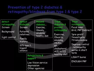
Management of patients with diabetic retinopathy • Prevention measures • Control of blood glucose levels • Control of blood pressure • Current treatment options • Retinal laser photocoagulation • Vitreoretinal surgery
Treatment: Laser Photocoagulation • Laser photocoagulation treatment can prevent vision loss in patients with PDR or DME • Several limitations • - Paliative, not curative • - Side-effect profile • ●Decreased night vision • ●Decreased peripheral vision
Accepted Goals for Medical Management of Diabetic Retinopathy Blood Glucose Control ● <7.0% HgA1C Blood Pressure Control ● <130/80 mms Hg LDL ● <100 mg/dl
D.R. PhenotypesRelevance for Clinical Management Different phenotypes – different rates of progression Phenotype A – Follow-up less frequent Phenotypes B and C – more frequent examinations.
D.R. PhenotypesRelevance for Clinical Management - To all: Tight metabolic control (HgA1C <7.1%) important - Special attention : Blood Pressure management – Phenotype B Coagulation cascade/platelets – Phenotype C
Prevention: Blood Glucose Control NHANES = National Health and Nutrition Examination Survey
Prevention: Blood Glucose Control JNC VII = 7th Report of the Joint National Committee on Prevention, Detection, Evalution and Treatment of High Blood Pressure.
Levels of therapy • 1st – Near physiological levels of glycemia • Initial stages of disease – • 2nd – Controlling biochemical events in the retina • - PKC inhibition • - AGE inhibition • - AR Inhibition • 3rd – Controlling initial vascular damage • - Blood-Retinal Barrier breakdown • - Anthithrombotic. Platelet inhibiton • - Antiinflammatory tx.
Targeted treatments for D.R. (initial stages) 1st component – similar to all phenotypes - controlling the biochemical damage of prolonged hyperglycemia - Intensive control of glycemic values - Inhibition of formation of advanced glycation end products - Inhibition of oxidative cellular stress-antioxidant drugs
Targeted treatments for D.R. (initial stages) 2nd component – depending on phenotypes Phenotype B – Modulation of the ability to regulate blood flow and leakage Phenotype C – Modulation of antithrombotic function of the endothelium
Targeted treatments for D.R. (initial stages) Phenotype B – Modulation of the ability to regulate blood flow and leakage - Inhibition of the diacylglycerol-protein kinase C pathway - Modulation of the nitric oxide pathway Phenotype C – Modulation of antithrombotic function of theendothelium - Platelet function inhibitors - Modulation of prostacyclin synthesis
– Summary – • Diabetes is highly prevalent and rapidly increasing worldwide • Diabetes retinopathy is a common microvascular complication of diabetes that affects the vision of 40% of inviduals with diabetes and threatens vision in about 8.2% of cases • Current therapy has limitations • Prevention of diabetic retinopathy with glycemic or blood pressure control is difficult to achieve in clinical practice • Laser therapy is effective but is associated with complications • Fundamental need for effective comunications between ophthalmologist and diabetologist
– Summary – - Diabetic Retinopathy - • Evaluate regularly since initial stages (NPDR). • Different Phenotypes. Different Rates of Progression. • Different risk profiles. • Different approaches to follow-up and treatment. • 5. Research on genotype-phenotype correlations.