Download
1 / 77
820 likes | 1.36k Vues
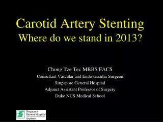
Carotid Artery Stenting Where do we stand in 2013?. Chong Tze Tec MBBS FACS Consultant Vascular and Endovascular Surgeon Singapore General Hospital Adjunct Assistant Professor of Surgery Duke NUS Medical School. Stroke. 3 rd leading cause of death in US

E N D
Carotid Artery StentingWhere do we stand in 2013? Chong Tze Tec MBBS FACS Consultant Vascular and Endovascular Surgeon Singapore General Hospital Adjunct Assistant Professor of Surgery Duke NUS Medical School
Stroke • 3rd leading cause of death in US • 750 000 people will have a stroke this year • 160 000 will die from it • 15-30% become permanently disabled • 20-30% caused by extracranial carotid disease
Carotid endarterectomy • Carotid artery stenting • Unresolved issues
CEA: Large-Scale Randomized Trials • ECST (1991) • NASCET (1991) • VA Asymptomatic Study (1993) • ACAS (1995) • ACST (2004)
NASCET 26% vs 9% rate at 2 years Barnett HJM et al NEJM 1998;339:1415-1425
ACAS 11% vs 5.1% rate at 5 years; p=.004 ACAS Investigators JAMA 1995;273:1421
Curves cross at 3 years Curves cross at 1.5 years
Carotid Stenting- Indications • Carotid restenosis • Anatomically difficult lesion (e.g. above C2) • Radiation-induced disease • “High-risk” patients - Consensus Conference, Montefiore Vascular Symposium 2001
Anatomic/technical Inaccessible lesion Hostile neck Radiation disease Restenotic lesion Comorbidities Age>80 CHF Recent coronary event or procedure COPD Contralateral occlusion Renal failure “High Risk” criteria for CEA ?
CEA vs CAS: Major RCTs • CAVATAS (Lancet 2001) • SAPPHIRE (NEJM 2004) • SPACE (Lancet 2006) • EVA-3S (NEJM 2006) • CREST (2010) • ICSS (2009)
SAPPHIRE Primary endpoints • Death/stroke/MI within 30 days • Death/ipsilateral stroke between 31 days and 1 year 747 patients were enrolled in the study and 334 patients underwent randomization. Of those not randomized, 406 entered into a stent registry and 7 entered a surgical registry
SAPPHIRE Discussion • CAS is not inferior to CEA in high risk patients based on 1 year data • Trial was terminated early due to the establishment of nonrandomized stent registries
SPACEStent-Supported Percutaneous Angioplasty of the Carotid Artery versus Endarterectomy
SPACE Hypothesis • CAS is not inferior to CEA for the treatment of severe symptomatic carotid stenosis
SPACE Discussion • SPACE failed to prove the non-inferiority of CAS compared to CEA • 30d stroke/death rate was 6.84% for CAS versus 6.34% for CEA • CEA 30d event rates are similar to NASCET (6.5%)
SPACE – Follow up results • Recurrent restenosis >70% was higher in the CAS group compared to the CEA group at 2 years • 10.7% vs 4.6%, p=0.0009
EVA-3S – Follow up results • Cumulative probability of periprocedural stroke or death and non-procedural ipsilateral stroke at 4 years
CAS vs CEA trials • Failed to show benefit so far • Perhaps there are subtleties involved which are underappreciated • Lesion characteristics • Technical aspects to CAS • Operator experience
Confounding issues • Arch anatomy • Stents design • Embolic protection devices • Plaque evaluation
Open cell stents are more conformable therefore offer better wall apposition and are more flexible and trackable
Cerebral embolization, as detected by TCD and DW-MRI, occurs with similar frequency After CAS with open-cell and closed-cell stents… does not support the superiority of any stent design with respect to cerebral embolization
Asymptomatic lesions (n=36) • Diffusion weighted MRI at 24h post procedure • Average number of hits 6.1 vs 6.2 • Filter group did not show reduction in microemboli