
Skeletal System
Skeletal System. FUNCTIONS OF SKELETON Support of trunk and skull Protection of underling organs Movement - attachment for muscles. Mineral homeostasis Hemopoiesis - all blood cells Energy Storage. CLASSIFICATION OF BONES Long bones Short bones Flat bones

Skeletal System
E N D
Presentation Transcript
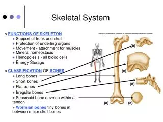
Skeletal System • FUNCTIONS OF SKELETON • Support of trunk and skull • Protection of underling organs • Movement - attachment for muscles • Mineral homeostasis • Hemopoiesis - all blood cells • Energy Storage • CLASSIFICATION OF BONES • Long bones • Short bones • Flat bones • Irregular bones • Seasmoid bone develop within a tendon • Wormian bones tiny bones in between major skull bones
Structure of Short, Irregular, and Flat Bones • Thin plates of periosteum-covered compact bone on the outside and endosteum-covered spongy bone within. • Have no diaphysis or epiphysis because they are not cylindrical. • Contain bone marrow between their trabeculae, but no marrow cavity. • In flat bones, the internal spongy bone layer is known as the diploë, and the whole arrangement resembles a stiffened sandwich.
Bone Marrow • Bone marrow is a general term for the soft tissue occupying the medullary cavity of a long bone, the spaces amid the trabeculae of spongy bone, and the larger haversian canals. • There are 2 main types: red & yellow. • Red bone marrow = blood cell forming tissue = hematopoietic tissue • Red bone marrow looks like blood but with a thicker consistency. • It consists of a delicate mesh of reticular tissue saturated with immature red blood cells and scattered adipocytes. Notice the red marrow and the compact bone
Distribution of Marrow Note the compact bone on the bottom and marrow on the bottom. • In a child, the medullary cavity of nearly every bone is filled with red bone marrow. • In young to middle-aged adults, the shafts of the long bones are filled with fatty yellow bone marrow. • Yellow marrow no longer produces blood, although in the event of severe or chronic anemia, it can transform back into red marrow • In adults, red marrow is limited to the axial skeleton, pectoral girdle, pelvic girdle, and proximal heads of the humerus and the femur.
Bone Classification 206 bones Axial skeleton • Forms long axis of the body. • Includes the bones of the skull, vertebral column, and rib cage. • These bones are involved in protection, support, and carrying other body parts. • Appendicular skeleton • Bones of upper & lower limbs and the girdles (shoulder bones and hip bones) that attach them to the axial skeleton. • Involved in locomotion and manipulation of the environment.
PARTS OF A LONG BONE • Diaphysis - shaft • Mainly compact bone • Medullary cavity • contains yellow marrow • lined with endosteum • Periosteum outer fibrous protective covering • osteogenic layer • attached to bone by Sharpey’s fibers • serves as an insertion for ligaments and tendons. • Epiphyses -proximal and distal ends • covered with articular cartilage • mainly spongy (cancellous) bone • trabeculae (small needle like projections) • red marrow-hematopoietic • contains the epiphyseal (growth) plate
Bone Features • Joint Structures • Fossa • Condyle • Epicondyle • Head • For Muscle attachment • Trochanter • Tuberosity • Process • Crest • Linea • Fovea • Tubercle • Passageways for vessels, etc. • Foramen • Fissure • Meatus • Space within a Bone - Sinus
Here, we see a cartoon showing all 3 cell types. Osteoblasts and osteoclasts are indicated. • Note the size of the osteoclast (compare it to the osteoblast), and note the ruffled border. • Why is there a depression underneath the osteoclast? • What is the name of the third cell type shown here? • What do you think the tan material represents?
The rod-shaped molecules lie in a staggered arrangement which acts as a template for bone mineralization. Bone mineral is laid down in the gaps. • Inorganic component of bone matrix • 2 salts: calcium phosphate and calcium hydroxide. • Interact to form a compound called hydroxyapatite. • Bone - magnesium, fluoride, and sodium. hardness resist compression. Collagen fibers cross section and occupy space btwn the black bone cells.
This bone: a. Has been demineralized b. Has had its organic component removed
MICROSCOPIC STRUCTURE • Spongy bone microscopic structure • trabeculae • spaces filled with red marrow • Compact bone Microscopic structure • Made of osteons (Haversian System) • concentric lamellae (calcified matrix) • central canal containing blood vessels and nerves • osteocytes in lacunae • Canals carrying nutrients to osteocytes • Canaliculi -connecting lacunae • Volkmans's -connecting osteons (right angles to central canals) • Chemical structure • Organic components (35%) • Cells • Osteoblasts • Osteocytes • Osteoclasts • Osteoid - mainly collagen for tensile strength • Inorganic component - 65% (hydroxyapatite- calcium phosphate) gives hardness to bone.
EmbryonicBone Development (Osteogeneis) fontanel • Intramembranous (ossification of fibrous CT) - flat bones • Osteoprogenitor cell differentiate into osteoblasts • Ossification's centers develop in the center of the bone • Periosteum develops and blood vessels invade developing centers • Endochondral(Ossification of hyaline cartilage) - long bones • Calcified cartilage • Ossification centers (primary & secondary) • Invasion of blood vessels • Formation of the medullary cavity • Epiphyseal plate
BONES OF SKULL, FACIAL BONES, CLAVIVLES, PELVIS, SCAPULAE, PART OF MANDIBLE
BONE GROWTHPhysical Stress – stimulates bone growth • Width (appositional growth)- occurs in nutrient periosteum • Bone remodeling - resorption and depositing • Length (longitudinal growth) - Epiphyseal plate • Proliferation zone • Zone of hypertrophy • Calcification of dead cells • Bone trabeculae --> Compact bone • Factors effecting bone growth/homeostasis • Nutrition - Ca, P, Mg, vitamins, A, C and D. • Hormones in general – Growth,Thyroid,Sex • Hormones that maintain blood calcium • Parathyroid - released when? • Vitamin D for Ca+ absorption • Resorption of Ca+ by kidneys • Stimulation of osteoclasts • Calcitonin - released when? • stimulates osteoblast activity
Growth in Bone Length • Epiphyseal cartilage (close to the epiphysis) of the epiphyseal plate divides to create more cartilage, while the diaphyseal cartilage (close to the diaphysis) of the epiphyseal plate is transformed into bone. This increases the length of the shaft.
At puberty, bone length is increased dramatically by activities of growth hormone thyroid hormone sex hormones. • As a result osteoblasts begin producing bone faster than the rate of epiphyseal cartilage expansion. • EPIPHYSEAL LINE
Bone Remodeling • Bone is a dynamic tissue. • What does that mean? • Wolff’s law holds that bone will grow or remodel in response to the forces or demands placed on it. Examine this with the bone on the left.
Why might you suspect someone whose been a powerlifter for 15 years to have heavy, massive bones, especially at the point of muscle insertion? • Astronauts tend to experience bone atrophy after they’re in space for an extended period of time. Why?
Bone Fractures Fracture Repair Osteoporosis Animation & another source


