Download
1 / 38
380 likes | 404 Vues
Learn why cells divide and the importance of cell division in growth, repair, and disease. Explore the stages of the cell cycle, DNA replication, and protein synthesis in an easy-to-understand format.

E N D
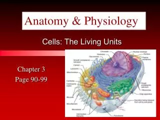
3 Cells: The Living Units Part D
Why Would a Cell Divide? As cells absorb nutrients and get larger, the volume of the cell increases faster than the surface area This means that a cell can no longer absorb nutrients and get rid of wastes fast enough to support its demands (volume) So what’s a cell to do? DIVIDE !!!!
Why Would a Cell Divide? Besides growth a cell would also divide for: Repair or Replacement Cancer Different cells divide at different rates: Most mammalian cells = 12-24 hours Some bacterial cells = 20-30 minutes
Getting Older… All cells are only allowed to complete a certain number of divisions Then they die (programmed cell death) How does cell division change over a lifetime? Childhood = cell division > cell death Adulthood = cell division = cell death The Later Years = cell division < cell death
Cell Cycle Tidbits How long is one cell cycle? • Depends on the cell- skin cells = ~24 hours, nerve cells = never after maturity, cancer cells = very short • Remember: every cell only has a certain # of divisions it can undergo, then it dies = apoptosis (programmed cell death)
Stages of the Cell Cycle There are two stages to a cells life. • interphase (growth & replication of DNA) • mitotic phase (division of cell into 2 daughter cells) Cell spends about 90% of the time in interphase
Interphase • Divided into 3 phases: • G1 (1st gap) = small cell is absorbing nutrients, growing & doing its job. • S (synthesis) = cell is continuing to grow & duplicates its DNA. • G2 (2nd gap) = cell keeps growing & doing its job.
Mitosis: A Closer Look • DNA is all twisted up into a Chromosome. • During S phase the Chromosome is copied. • 2 complete identical sets of chromosomes. • They are connected in the middle by a centromere. • A single copied chromosome is called a Chromatid.
DNA Replication • DNA helices begin unwinding from the nucleosomes • Helicase untwists the double helix and exposes complementary strands • The site of replication is the replication bubble • Each nucleotide strand serves as a template for building a new complementary strand
DNA Replication • The replisome uses RNA primers to begin DNA synthesis • DNA polymerase III continues from the primer and covalently adds complementary nucleotides to the template PLAY DNA Replication
DNA Replication • Since DNA polymerase only works in one direction: • A continuous leading strand is synthesized • A discontinuous lagging strand is synthesized • DNA ligase splices together the short segments of the discontinuous strand • Two new telomeres are also synthesized • This process is called semiconservative replication
DNA Replication Figure 3.31
Cell Division • Essential for body growth and tissue repair • Mitosis – nuclear division • Cytokinesis – division of the cytoplasm
The Mitotic Phase • Divided into 4 stages of Mitosis: • Prophase • Metaphase • Anaphase • Telophase (+) PLUS • Cytokinesis
Early and Late Prophase • Asters are seen as chromatin condenses into chromosomes • Nucleoli disappear • Centriole pairs separate and the mitotic spindle is formed PLAY Prophase PLAY Prometaphase
Early Prophase Pair of centrioles Early mitotic spindle Centromere Aster Chromosome, consisting of two sister chromatids Early prophase Figure 3.32.2
Late Prophase Fragments of nuclear envelope Polar microtubules Kinetochore Kinetochore microtubule Spindle pole Late prophase Figure 3.32.2
Metaphase • Chromosomes cluster at the middle of the cell with their centromeres aligned at the exact center, or equator, of the cell • This arrangement of chromosomes along a plane midway between the poles is called the metaphase plate PLAY Metaphase
Metaphase Metaphase Figure 3.32.4
Anaphase • Centromeres of the chromosomes split • Motor proteins in kinetochores pull chromosomes toward poles PLAY Anaphase
Anaphase Daughter chromosomes Anaphase Figure 3.32.5
Telophase and Cytokinesis • New sets of chromosomes extend into chromatin • New nuclear membrane is formed from the rough ER • Nucleoli reappear • Generally cytokinesis completes cell division PLAY Telophase
Telophase and Cytokinesis Nucleolus forming Contractile ring at cleavage furrow Nuclear envelope forming Telophase and cytokinesis Figure 3.32.5
Cytokinesis • Cleavage furrow formed in late anaphase by contractile ring • Cytoplasm is pinched into two parts after mitosis ends
Protein Synthesis • DNA serves as master blueprint for protein synthesis • Genes are segments of DNA carrying instructions for a polypeptide chain • Triplets of nucleotide bases form the genetic library • Each triplet specifies coding for an amino acid
From DNA to Protein Figure 3.34
Roles of the Three Types of RNA • Messenger RNA (mRNA) carries the genetic information from DNA in the nucleus to the ribosomes in the cytoplasm • Transfer RNAs (tRNAs) bound to amino acids base pair with the codons of mRNA at the ribosome to begin the process of protein synthesis • Ribosomal RNA (rRNA) is a structural component of ribosomes
Transcription • Transfer of information from the sense strand of DNA to RNA • Transcription factor • Loosens histones from DNA in the area to be transcribed • Binds to promoter, a DNA sequence specifying the start site of RNA synthesis • Mediates the binding of RNA polymerase to promoter
Transcription: RNA Polymerase • An enzyme that oversees the synthesis of RNA • Unwinds the DNA template • Adds complementary ribonucleoside triphosphates on the DNA template • Joins these RNA nucleotides together • Encodes a termination signal to stop transcription
Overview of Transcription Figure 3.35
Initiation of Translation • A leader sequence on mRNA attaches to the small subunit of the ribosome • Methionine-charged initiator tRNA binds to the small subunit • The large ribosomal unit now binds to this complex forming a functional ribosome
Polypeptide Chain Elongation Figure 3.37
Genetic Code • RNA codons code for amino acids according to a genetic code Figure 3.36
Information Transfer from DNA to RNA • DNA triplets are transcribed into mRNA codons by RNA polymerase • Codons base pair with tRNA anticodons at the ribosomes • Amino acids are peptide bonded at the ribosomes to form polypeptide chains • Start and stop codons are used in initiating and ending translation
Information Transfer from DNA to RNA Figure 3.39
Developmental Aspects of Cells • All cells of the body contain the same DNA but develop into all the specialized cells of the body • Cells in various parts of the embryo are exposed to different chemical signals that channel them into specific developmental pathways • Genes of specific cells are turned on or off • Cell specialization is determined by the kind of proteins that are made in that cell
Developmental Aspects of Cells • Development of specific and distinctive features in cells is called cell differentiation • Cell aging • Wear and tear theory attributes aging to little chemical insults and formation of free radicals that have cumulative effects throughout life • Genetic theory attributes aging to cessation of mitosis that is programmed into our genes