Skin and Its Layers
540 likes | 573 Vues
Learn about the structure of the skin, its layers (epidermis, dermis, hypodermis), cells, growth factors, junctions, and functions. Discover how skin color is determined by melanin types and factors affecting it.

Skin and Its Layers
E N D
Presentation Transcript
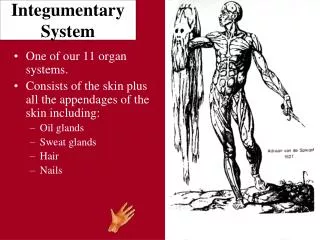
INTRODUCTION Skin (integument) is the body’s largest organ Approximately 1.6 to 1.9 m2 in average-sized adult Integumentary system describes the skin and its appendages: hair, nails, and skin glands
STRUCTURE OF THE SKIN Skin classified as a cutaneous membrane Two primary layers: epidermis and dermis; joined by dermoepidermal junction (Figures 6-1 and 6-2) Hypodermis lies beneath dermis Thin and thick skin (Figure 6-3) Thin skin covers most of the body’s surface (1 to 3 mm thick) Thick skin covers soles and palms (4 to 5 mm thick)
STRUCTURE OF THE SKIN: EPIDERMIS Epidermis Cell types (Figure 6-4) Keratinocytes: constitute more than 90% of cells present; principal structural element of the outer skin; sometimes called corneocytes after they are fully keratinized Melanocytes: pigment-producing cells (5% of the total); contribute to skin color and filter ultraviolet (UV) light Epidermal dendritic cells: branched antigen-presenting cells; play a role in immune response; also called Langerhans cells Tactile epithelial cells (Merkel cells): attach to sensory nerve endings to form “light touch” receptors
STRUCTURE OF THE SKIN: EPIDERMIS (cont.) Cell layers Stratum basale (base layer): single layer of columnar cells; only these cells undergo mitosis and then migrate through the other layers until they are shed Stratum spinosum (spiny layer): cells arranged in eight to 10 layers with desmosomes that pull cells into spiny shapes; cells rich in RNA Stratum germinativum (growth layer): another name for stratum basale or stratum spinosum and stratum basale together
STRUCTURE OF THE SKIN: EPIDERMIS (cont.) Stratum granulosum (granular layer): cells arranged in two to four layers and filled with keratohyalin granules; contains high levels of lysosomal enzymes Stratum lucidum (clear layer): cells filled with keratin precursor called eleidin; absent in thin skin Stratum corneum (spiked layer): most superficial layer; dead cells filled with keratin (barrier area)
Epidermal growth and repair Turnover or regeneration time refers to time required for epidermal cells to form in the stratum basale and migrate to the skin surface—approximately 35 days Epidermal growth factor regulates epidermal growth and repair Shortened turnover time will increase the thickness of the stratum corneum and result in callus formation Normally 10% to 12% of all cells in stratum basale enter mitosis daily Each group of eight to 10 basal cells in mitosis with their vertical columns of migrating keratinocytes is called an epidermal proliferating unit STRUCTURE OF THE SKIN: EPIDERMIS (cont.)
STRUCTURE OF THE SKIN: EPIDERMIS (cont.) Dermoepidermal junction A basement membrane with unique fibrous elements and a polysaccharide gel “glue” the epidermis to the dermis below The junction is a partial barrier to the passage of some cells and large molecules
STRUCTURE OF THE SKIN: DERMIS Dermis Sometimes called “true skin”—much thicker than the epidermis and lies beneath it Gives strength to the skin Serves as a reservoir storage area for water and electrolytes Contains various structures Arrector pili muscles and hair follicles (Figure 6-5) Sensory receptors (Figure 6-6) Sweat and sebaceous glands Blood vessels Rich vascular supply plays a critical role in temperature regulation
STRUCTURE OF THE SKIN: DERMIS (cont.) Layers of dermis Papillary layer: composed of dermal papillae that project into the epidermis; contains fine collagenous and elastic fibers and the dermoepidermal junction; forms a unique pattern that gives individual fingerprints Reticular layer: contains dense, interlacing white collagenous fibers and elastic fibers to make the skin tough yet stretchable; when processed from animal skin, produces leather
STRUCTURE OF THE SKIN: DERMIS (cont.) Dermal growth and repair The dermis does not continually shed and regenerate itself as does the epidermis During wound healing, fibroblasts begin forming an unusually dense mass of new connective fibers; if not replaced by normal tissue, this mass remains a scar Cleavage lines: patterns formed by the collagenous fibers of the reticular layer of the dermis; also called Langer’s lines (Figure 6-7)
STRUCTURE OF THE SKIN: HYPODERMIS Hypodermis Also called the subcutaneous layer or superficialfascia Located deep to the dermis; forms connection between skin and other structures Not part of the skin
SKIN COLOR Melanin Basic determinant is quantity, type, and distribution of melanin Types of melanin Eumelanin: group of dark brown, almost black, melanins Pheomelanin: group of reddish and orange melanins Melanin formed from tyrosine by melanocytes (Figure 6-8) Melanocytes release melanin in packets called melanosomes Melanosomes are ingested by surrounding keratinocytes and form a cap over the nucleus
SKIN COLOR (cont.) Albinism: congenital absence of melanin Process regulated by tyrosinase, exposure to sunlight (UV radiation), and certain hormones, including melanocortins (adrenocorticotropic hormone, alpha melanocyte stimulating hormone) and endothelin-1 (Figures 6-9 and 6-10) Cumulative effects of UV ray exposure may produce age spots (Figure 6-11)
SKIN COLOR: OTHER PIGMENTS Other pigments Beta-carotene (group of yellowish pigments from food) can also contribute to skin color Hemoglobin: color changes also occur as a result of changes in blood flow Redder skin color when blood flow to skin increases Cyanosis: bluish color caused by darkening of hemoglobin when it loses oxygen and gains carbon dioxide (Figure 6-12) Bruising can cause a rainbow of different colors to appear in the skin (Figure 6-13) Other pigments: from cosmetics, tattoos, and bile pigments in jaundice (Box 6-4)
FUNCTIONS OF THE SKIN Protection (Table 6-2) Physical barrier to microorganisms Barrier to chemical hazards Reduces potential for mechanical trauma Prevents dehydration Protects from excess UV ray exposure (melanin function)
FUNCTIONS OF THE SKIN (cont.) Surface film Emulsified protective barrier formed by mixing of residue and secretions of sweat and sebaceous glands with sloughed epithelial cells from skin surface; shedding of epithelial elements is called desquamation Functions Antibacterial, antifungal activity Lubrication Hydration of skin surface Buffer of caustic irritants Blockade of toxic agents
FUNCTIONS OF THE SKIN (cont.) Chemical composition From epithelial elements: amino acids, sterols, and complex phospholipids From sebum: fatty acids, triglycerides, and waxes From sweat: water, ammonia, urea, and lactic and uric acid
FUNCTIONS OF THE SKIN (cont.) Sensation Skin acts as a sophisticated sense organ Somatic sensory receptors detect stimuli that detection of pressure, touch, temperature, pain, and other general senses Flexibility Skin is supple and elastic, thus permitting change in body contours without injury
FUNCTIONS OF THE SKIN (cont.) Excretion Water Urea/ammonia/uric acid Hormone (vitamin D) production (Figure 6-14) Exposure of skin to UV light converts 7-dehydrocholesterol to cholecalciferol, a precursor to vitamin D Blood transports precursor to liver and kidneys, where vitamin D is produced Process and end result fulfill the necessary steps required for vitamin D to be classified as a hormone
FUNCTIONS OF THE SKIN (cont.) Immunity Phagocytic cells destroy bacteria Epidermal dendritic cells trigger helpful immune reaction working with helper T cells Homeostasis of body temperature To maintain homeostasis of body temperature, heat production must equal heat loss; skin plays a critical role in this process Heat production By metabolism of foods in skeletal muscles and liver Chief determinant of heat production is the amount of muscular work being performed
FUNCTIONS OF THE SKIN (cont.) Homeostasis of body temperature Heat loss: approximately 80% of heat loss occurs through the skin; remaining 20% occurs through the mucosa of the respiratory, digestive, and urinary tracts (Figure 6-15) Evaporation: to evaporate any fluid, heat energy must be expended; this method of heat loss is especially important at high environmental temperatures when it is the only method heat can be lost from the skin Radiation: transfer of heat from one object to another without actual contact; important method of heat loss in cool environmental temperatures Conduction: transfer of heat to any substance in contact with the body; accounts for relatively small amounts of heat loss Convection: transfer of heat away from a surface by movement of air; usually accounts for a small amount of heat loss
FUNCTIONS OF THE SKIN (cont.) Homeostatic regulation of heat loss (Figure 6-16) Heat loss by the skin is controlled by a negative feedback loop Receptors in the hypothalamus monitor the body’s internal temperature If body temperature is increased, the hypothalamus sends a nervous signal to the sweat glands and blood vessels of the skin The hypothalamus continues to act until the body’s temperature returns to normal
APPENDAGES OF THE SKIN Hair (Figure 6-17) Development of hair Distribution is over entire body except palms of hands and soles of feet and a few other small areas Fine and soft hair coat present before birth called lanugo Coarse pubic and axillary hair that develops at puberty called terminal hair Hair follicles and hair develop from epidermis; mitosis of cells of germinal matrix forms hairs Papilla: cluster of capillaries under germinal matrix Root: part of hair embedded in follicle in dermis Shaft: visible part of hair Medulla: inner core of hair Cortex: outer portion
APPENDAGES OF THE SKIN: HAIR (cont.) Appearance of hair Color: result of different amounts, distribution, types of melanin in cortex of hair (Figure 6-18) Growth: growth and rest periods alternate; hair on head averages 5 inches of growth per year Sebaceous glands attach to and secrete sebum (skin oil) into follicle Male pattern baldness (androgenic alopecia) results from combination of genetic tendency and male sex hormones (Figure 6-19)
APPENDAGES OF THE SKIN (cont.) Nails (Figure 6-20) Consist of epidermal cells converted to hard keratin Nail body: visible part of each nail Root: part of nail in groove hidden by fold of skin, the cuticle Lunula: moon-shaped white area nearest root Nail bed: layer of epithelium under nail body; contains abundant blood vessels Appears pink under translucent nails Nails may have pigmented streaks (Figure 6-21) Separation of a nail from the nail bed is called onycholysis (Figure 6-22) Growth: nails grow by mitosis of cells in stratum basale beneath the lunula; average growth about 0.5 mm/week, or slightly over 1 inch per year
APPENDAGES OF THE SKIN (cont.) Skin glands (Figure 6-23) Two types of sweat glands Eccrine glands Most numerous sweat glands; quite small Distributed over total body surface with exception of a few small areas Simple, coiled, tubular glands Function throughout life Secrete perspiration or sweat; eliminate wastes and help maintain a constant core temperature Apocrine glands Located deep in subcutaneous layer Limited distribution: axilla, areola of breast, and around anus Large (often more than 5 mm in diameter) Simple, branched, tubular glands Begin to function at puberty Secretion shows cyclic changes in female with menstrual cycle