Download
1 / 6
60 likes | 215 Vues
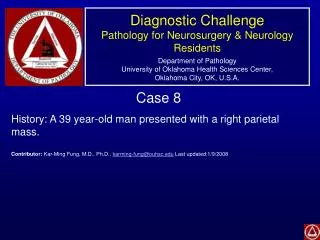
Diagnostic Challenge Pathology for Neurosurgery & Neurology Residents Department of Pathology University of Oklahoma Health Sciences Center, Oklahoma City, OK, U.S.A. Case 8 History: A 39 year-old man presented with a right parietal mass.

E N D
Diagnostic ChallengePathology for Neurosurgery & Neurology ResidentsDepartment of PathologyUniversity of Oklahoma Health Sciences Center,Oklahoma City, OK, U.S.A. Case 8 History: A 39 year-old man presented with a right parietal mass. Contributor: Kar-Ming Fung, M.D., Ph.D., karming-fung@ouhsc.edu Last updated:1/9/2008
Paraffin Section-Hematoxylin & Eosin (HE) Paraffin Section-Luxol Fast Blue-Periodic Acid Schiff (LFB-PAS) A B
HE LFB-PAS C D
CD163 (macrophages) GFAP (astrocytes) F E Neurofilament (axons) LCA (lymphocytes) H G
Diagnosis: Demyelinating process. • Discussion: • On scanning magnification, there are areas of well defined increased histology and these areas loss the “pinkness” of brain parenchymal tissue. On LFB-PAS stain, these areas does not contain myelin. (Panel A and B) Luxol fast blue is a stain that could highlight myelin. • On high magnification, the areas with increased cellularity has many foamy macrophages (arrow in the panel below). (Panel C and D). • On immunohistochemistry, the area with increased cellularity has numerous macrophages (CD68) and reactive, stellate shaped astrocytes (GFAP). (Panel E and F). • The axons are well preserved as shown by immunohistochemistry for neurofilament. (Panel G) • Very little lymphocytes are present (Panel H). The number of lymphocytes in a demyelinating process is usually quite variable. • There are no atypical cells or necrosis to suggest progressive multifocal leukoencephalopathy (PML). • Important message: It is important to realize that the high cellularity is not resulted from neoplastic changes. Demyelinating processes can be easily misdiagnosed as neoplastic changes and may potentially lead to disastrous consequences.