Download
1 / 10
100 likes | 237 Vues
Magnetic R esonance Imaging -THE MRI- . By: Megan Johnson Technology and Society Final Exam. Inventors of the MRI:. In 1924, Wolfgang Pauli – atoms have magnetic properties--basis of the NMR theory In1938, I. I. Rabi- first NMR device (substance’s magnetic properties)

E N D
Magnetic Resonance Imaging-THE MRI- By: Megan Johnson Technology and Society Final Exam
Inventors of the MRI: • In 1924, Wolfgang Pauli – atoms have magnetic properties--basis of the NMR theory • In1938, I. I. Rabi- first NMR device (substance’s magnetic properties) • In 1945, Physicists at Harvard University Felix Bloch and Edward Purcell - also created a NMR device (that could provide information about many types of classifications with no limitations)
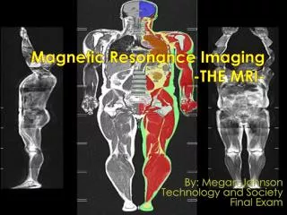
MRI of the abdomen (kidneys and liver) MRI of the Knee
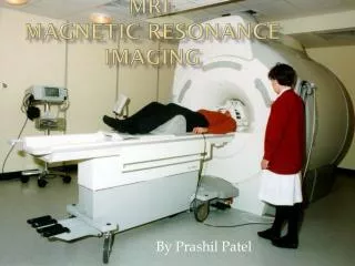
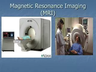
How all MRI’s are used: • In a special MRI suite, the patient lies down on a narrow table. • Transmitters are positioned on the body and the table moves into a long tube that houses the magnet. • Once the area to be examined has been properly positioned, a radio pulse is applied. Then a two-dimensional image corresponding to one slice through the area is made. The table then moves a fraction of an inch and the next image is made. Each image exposure takes several seconds and the entire exam will last anywhere from 30 to 90 minutes. (must stay still) • Depending on the area to be imaged, the radio-wave transmitters will be positioned in different locations. • For the head and neck, a helmet-like covering is worn on the head. • For the spine, chest, and abdomen, the patient will be lying on the transmitters. • For the knee, shoulder, or other joint, the transmitters will be applied directly to the joint. • Also during the test, Additional probes will monitor vital signs.
Abnormalities which MRI can find: • An organ is too large, too small, damaged, or absent. • Abnormal growths (such as tumors) are present. • Abnormal fluid from a cause such as bleeding or an infection is present. Fluid is found around the lungs or heart. Fluid is found around the liver, bowel, or other organ in the abdomen. • A blood vessel is narrowed or blocked. An aneurysm is present. • Blockage in the gallbladder bile ducts or in the tubes that lead out of the kidneys is present. • Damage to Joints, ligaments, or cartilage is seen. Bones are broken or show infection or disease. • Problems of the nervous system are present, such as multiple sclerosis (MS), dementia, Alzheimer's disease, or herniated disc.
POSTIVES of the MRI • precise images • This device shows tissues deep within the body or even the surface, also finding diseased tissue that are still in their first stages. • The Modern MRI uses cross-sectional, from one side of body to another, or three-dimensional images. • The MRI are very helpful when a surgery needs to be preformed. • There are no physical side effects or health risks that eligible patients have to worry about.
Negatives of the MRI • false positives due to the fact that they have herniated or bulging discs. • These people have no clinical symptoms that suggest a problem that the MRI is suggesting. • Along with false positives there are some restrictions with patient’s ability to use this machine. • Claustrophobic • Ferromagnetic devices (metallic iron) • Poor quality of MRI images comes from movement within the scanner • The patient needs to remain as still a possible for 30 to 90 minutes that the imaging last • Also many patients wonder about unborn babies, this is a discussion that needs to be discussed with the women’s doctor because it varies with each patient.
Cultural and Societal effects: • The advancements of the MRI have really set precedence among the medical world, with the availability of this Machine increasing more and more patients have the option of the MRI. • Save lives that without these technologies would have been lost • We have grown to have a dependence on medication, surgeries, therapy, etc for cures. – Medical advances in general • We now have more people living to see 100 years old • People are being diagnosed with cancer and overcoming it • Simple medications to prevent colds, and there are so many more technologies that have truly changed our world • The environment is not directly affected by my invention but it does indirectly. • The ability to diagnosis patients and prevent future health problems has helped our society to grow therefore causing expansion: more buildings, more families, more kids, and simply more on the earth. • I believe there will be a push for machine that can create images such as the MRI and CAT scan that do not have magnets or radiation side effects. • There is hope for the future of diagnosis tests to be capable of more than just images but maybe even being able to tell the problem or treat it.