
Specific Aims
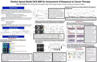
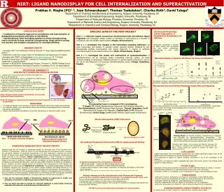
Shutter-Speed Model DCE-MRI for Assessment of Response to Cancer Therapy U01 CA154602; Wei Huang, PhD, Christopher Ryan, MD; Oregon Health & Science University, Portland, Oregon. Evaluation of Residual Cancer Burden (RCB) after Neoadjuvant Chemotherapy. Specific Aims.

Specific Aims
E N D
Presentation Transcript
Shutter-Speed Model DCE-MRI for Assessment of Response to Cancer Therapy U01 CA154602; Wei Huang, PhD, Christopher Ryan, MD;Oregon Health & Science University, Portland, Oregon • Evaluation of Residual Cancer Burden (RCB) after Neoadjuvant Chemotherapy Specific Aims Scatter plots of % changes at TP1 (relative to TP0) in RECIST, ROI ADC, ROI and histogram median ΔKtrans values vs. % necrosis at surgery for 9 sarcoma patients. There were significant linear correlations between % necrosis and % changes in ROI ΔKtrans (R = -0.93, P = 0.0003; Spearman’s correlation), and histogram median ΔKtrans (R = -0.71, P = 0.03). • Specific Aim 1: Compare Shutter-Speed model (SSM) DCE-MRI with standard model (SM) DCE-MRI, diffusion-weighted (DW) MRI, and tumor size for early prediction of therapy response and accurate assessment of post-therapy residual cancer. • Breast cancer neoadjuvant chemotherapy • Soft tissue sarcoma phase I/II trial with Sorafenib • Correlation with pathology endpoints • Specific Aim 2: Investigate the effects of data acquisition and processing schemes on DCE-MRI biomarkers for the purpose of assessing therapy response. • Temporal resolution • Acquisition time • AIF variation • Specific Aim 3: Develop software tools that can provide clinicians with a single and/or a set of biomarker values to aid clinical decision-making. Significant correlations between residual cancer burden (RCB) and MRI metrics measured after NACT completion (V4, or TP3, before surgery). • Evidence for Caution in DCE-MRI Assessment of Response to Antiangiogenic Treatment Using Reference Tissue Method Specific Aim 2 • Effects of Acquisition Time on DCE-MRI Assessment of Breast Cancer Response to Neoadjuvant Chemotherapy Column graphs of (a) TP1 tumor mean Ktrans (SM and SSM) and kep (SM and SSM) values and (b) their corresponding % changes (TP1 relative to TP0) at simulated, varying DCE-MRI acquisition time (Tacq). The blue column represents the mean pCR value, while the red column represents the mean non-pCR value. The results suggest that for typical responder and non-responder breast tumor Ktrans or kep values and their changes after one chemotherapy cycle, a DCE-MRI Tacq of 5-6 min is sufficient to achieve the goal of early prediction of therapeutic response when either parameter is used as the discriminatory biomarker. Study Schema Left: SM and SSM Ktrans maps of normal appearing muscle ROIs adjacent to the sarcoma of one patient at TP0 and TP1. Right: the decreases of SM and SSM muscle Ktrans in eight sarcoma patients from TP0 to TP1 were statistically significant: paired t test, P < 0.05. Progress Report Specific Aim 1 • Early Prediction of Soft-Tissue Sarcoma Response to Antiangiogenic Therapy • Early Prediction of Breast Cancer Response to Neoadjuvant Chemotherapy • Effects of Model/Algorithm on DCE-MRI Assessment of Breast Cancer Response to Neoadjuvant Chemotherapy • See DCE-/DW-MRI WG poster • Image Analysis Pipeline and Workflow. • The OHSU informatics group is working on several tasks pertaining to Specific Aim 3: • Modeling existing imaging workflow at OHSU Advanced Imaging Research Center (AIRC) and draft design of new, integrated, workflow (Image Analysis Pipeline). • Beginning procurement and setup up computing environment for Image Analysis Pipeline. • Beginning process to install and test open source software needed for Image Analysis Pipeline. • Establish a project management framework (project plan/tasks, communication plan, deliverables and timeline). • Submitting images to the central TCIA repository. • Optimizing a pipeline for image registration across time (longitudinal) and image type (T1-weighted DCE-MRI and parametric maps overlaid on anatomical images). • Optimizing a pipeline for motion correction using open-source algorithms (FLIRT, FNIRT, ANTS, BrainsFit are being evaluated currently). • Modifying the user-interface for XNAT to incorporate a clinician centric view. • Begin development of a DCE-MRI data analysis module in 3D Slicer. • Begin a clinical needs assessment with clinicians. Specific Aim 3 Ktrans (SSM), Δktrans, and τi maps of two breast tumors before neoadjuvant chemotherapy (NACT) (V1, or TP0) and after one NACT cycle (V2, or TP1). One is a pathologic complete responder (pCR, bottom two rows) and the other is a pathologic partial responder (pPR, top two rows) – non-pCR. Response status was determined by surgical pathology after 6 NACT cycles. Column graph of mean % changes of MRI metrics after one NACT cycle for the pCR (n = 4) and the non-pCR (n = 11) groups. The error bar represents the SD value. Except for RECIST, the other DCE-MRI metrics are excellent early predictors of response. SM, SSM Ktrans and ΔKtrans maps of two sarcomas at time point zero – baseline (TP0) and TP1 (after two weeks of antiangiogenic therapy only). Pathology reviews of surgical specimens at TP2 revealed that one tumor had optimal response to treatment (>95% necrosis) (top panels), while the other had sub-optimal response (50% necrosis) (bottom panels).


![Specific Aims Workshop [NAME ] [“TITLE”]](https://cdn5.slideserve.com/9671983/specific-aims-workshop-name-title-dt.jpg)