Download
1 / 1
10 likes | 175 Vues
FNf-ANP 50 nm. FNf-ANP 30 nm. Fragment. Migration ← Cdc42, Rac ← JNK. ERK → MLCK → Migration. Ligand Binding. FNf-ANP 100 nm. FNf-ANP 75 nm. FNf. Ligand-Nanoparticle Trafficking Aim 3 (Talaga, Co- PI). Cell Motility Dynamics Aim 1 (Moghe, PI). Trafficking.

E N D
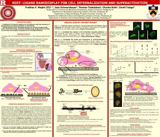
FNf-ANP 50 nm FNf-ANP 30 nm Fragment Migration ← Cdc42, Rac← JNK ERK →MLCK → Migration Ligand Binding FNf-ANP 100 nm FNf-ANP 75 nm FNf Ligand-Nanoparticle Trafficking Aim 3 (Talaga, Co- PI) Cell Motility Dynamics Aim 1 (Moghe, PI) Trafficking Nanoscale Biosensing of Cytointernalization Aim 3 (Tsakolakos, Co- PI) Internalization Signaling Aim 2 (Roth, Co- PI) (Gene) (Schwarzbauer, Co-PI, Moghe, PI) (Protein) Migration ← Cdc42, Rac← JNK ERK →MLCK → Migration Ligand Binding Ligand Design Aim 1 (Schwarzbauer, Co-PI) Nanoparticle Design Aim 1 (Moghe, PI) Fn I 1-5 Fibrin Heparin Synergy FnIII12-14 RGD FnI10-12 Heparin Fibrin NH2 COOH S S Gelatin Collagen Cell Binding Domain Variant Mix Fn III 9-10 Temperature and pH treatment Post-Internalization Processing Separate Integrins Integrin Recycling To leading edge of cell Albumin Denatured albumin Lysosome Trafficking To lysosome Internalization Clathrin Mediated Albumin Nanoparticle (ANP) Cytoskeletal Rearrangements Cdc42, Rac NIRT: LIGAND NANODISPLAY FOR CELL INTERNALIZATION AND SUPERACTIVATION Prabhas V. Moghe (PI)1, 2, Jean Schwarzbauer3, Thomas Tsakalakos4, Charles Roth2, David Talaga5 1Department of Chemical and Biochemical Engineering, Rutgers University, Piscataway, NJ 2Department of Biomedical Engineering, Rutgers University, Piscataway, NJ 3Department of Molecular Biology, Princeton University, Princeton, NJ 4Department of Materials Science and Engineering, Rutgers University, Piscataway, NJ 5Department of Chemistry and Chemical Biology, Rutgers University, Piscataway, NJ INTELLECTUAL MERIT SPECIFIC AIMS OF THE NIRT PROJECT Aim 1 is to fabricate organic nanocarriers functionalized with cell-adhesive ligand fragments and investigate cellular motility on interfaces of nanocarriers as a function of nanocarrier size and variable ligand loading. (Moghe, Schwarzbauer) Aim 2 is to investigate the changes in the intracellular signaling pathways (via gene microarrays and studies of selected protein signaling targets) activated by cell-internalizable particles functionalized with ligand fragments in relation to substrate immobilized ligand-functionalized nanocarriers. (Roth, Schwarzbauer, Moghe) Aim 3 is to investigate the modes and mechanisms of cytointernalization of ligandnanocarriers via (a) molecular studies modulating endocytic uptake; (b) single molecule imaging of intracellular trafficking kinetics of reporter ligand nanocarriers; and (c) the use of new biosensors for nanoscale cytointernalization events. (Talaga, Tsakalakos, Moghe) Keratinocyte Morphology F-Actin Organization is Modulated by Nanodisplay and ANP Size -- Combination of Dynamic Approach to Cell Adhesion and Internalization of Nanoparticles Accelerates Activation of Cell Motility -- Interdisciplinary Team leveraging expertise in cellular bioengineering, matrix molecular biology, molecular bioengineering, single particle tracking and analysis, and inorganic nanoparticles for sensing/trafficking Keratinocyte morphology is affected by ligand display and nanoparticle size, with smaller nanoparticles demonstrating a more motile phenotype. BROADER IMPACTS • -- Synergies with T32 Postdoctoral Training Program on Tissue Engineering/Biomaterials Science • --Collaboration with New Jersey Center for Biomaterials and Department of Defense • --Synergies with IGERT on Biointerfaces (www.igert.rutgers.edu, PI: P. Moghe) • Diversity Infrastructure: NE-AGEP; Institute for Teaching & Mentoring; • Leadership in Diversity Award-- • Outreach to RISE/ISURF Undergraduate Program. Princeton U., RWJMS Medical School • --Translational Potential for Anti-Aging Therapies; Foundations for Ctr for Nanomedicine Nanoscale Display in Dynamic Interfaces Increases Cell Migration • MOTIVATION: BARRIERS TO • SKIN MOTILITY DURING RE-EPITHELIALIZATION • • Largest organ in the human body; protects it from water loss, • heat, and microorganisms • • For proper wound healing, need to re-establish the epidermal layer • via skin cell migration to close the wound site • • Defects in re-epithelialization processes can to lead deficient • Healing, chronic wounds, or in extreme cases morbidity and mortality • • Chronic wounds cost about $3 billion annually • • Diabetic foot ulcers affect 1 million in the U.S. (86,000 amputations) • • Manipulation of matrix components can be used to modify cellular • responses to improve wound repair • GOAL: Enhance cellular activity and dynamics via nanoscale • engineered ligand presentation • HOW: Nanodisplay of engineered biorelevant fibronectin fragments to cells via albumin nanoparticles • HYPOTHESES • The display of ligands on the nanoscale via albumin nanoparticles will promote integrin nanoclustering. • The substrate mobility of the nanoparticles will promote membrane-based ligand/integrin translocation and possible internalization. • Integrin nanoclustering & internalization will activate cell signaling for enhanced cellular motility. Cell migration kinetics is sensitively governed by ligand concentration, nanocarrier size, mobility and uptake mechanisms, suggesting that nanoparticles must be internalized by cells to induce cell superactivation. Nanoparticle Size and Mobility Modulates Uptake Rate and Signaling The assembled team is composed of five investigators: P. Moghe, PI (Cellular Bioengineering; Nanoscale Cell-Interactive Materials); Jean Schwarzbauer, Co-PI (Molecular Matrix Biology); Thomas Tsakalakos, Co-PI (Nanostructured Inorganic Bio-Materials); Charles Roth, Co-PI (Molecular Bioengineering); David Talaga, Co-PI (Single Molecule Spectroscopy & Biophysics) Smaller sized nanoparticles can be taken up at faster rates than larger sized nanoparticles. Nanoparticle size and mobility can amplify relevant signaling element expression, which could be leading to superactivation of cell motility. 5 integrin Green 1 integrin Red PBS vs. Albumin Nanoparticle (ANP) Fabrication Integrin Ligand Nanocarrier Whole length Fibronectin Thinking Model: On dynamic interfaces, keratinocytes can sequester and internalize the ligand-functionalized nanoparticles. The internalization increases with smaller sized carriers, leading to increased rates of post-uptake processing and signaling within the cell. Nanodisplayed ligand Highly clustered integrins & Cytointernalizable Carriers Ligand adsorbed surfaces Non - or slightly clustered integrins FNf-ANP Recombinant Fragment of Fibronectin (FNf) for Engineered Cell Motility Preliminary data: Integrin a5b1 clustering and diffusivity throughout the cell membrane is enhanced when keratinocytes are presented with FNf-ANP’s. Further studies are underway to examine the integrin and endosomal dynamics. • OVERVIEW & ORGANIZATION OF THE NIRT PROJECT • Nanotechnology has the potential to significantly amplify the functional roles of the currently known repertoire of biomolecules. While the search for new biomolecules with therapeutic function continues, the cell-specific exposure of established biomolecules is far from optimized to maximize the cellular function of interest. Since cellular binding and subsequent interactions with extracellular biomolecules are events occurring at the nanoscale, cellular engineering affords a fertile landscape for controlled application of nanotechnology. To successfully exploit such opportunities, however, will require integrative studies of the roles of molecules in dynamic systems. At Rutgers, we have discovered that fragments of cell adhesion biomolecules like fibronectin, which has been widely studied for its effects on cell adhesion, growth, migration, can elicit markedly altered cell responses, with consequences for internalization and superactivation of cell functions, if displayed on a nanoscale. The substrate-based display of fragments of adhesion molecules from nanoscale carriers affects receptor-mediated cell binding followed by the internalization of the adhesion ligands. The concerted remodeling and internalization processes appear to superactivate intracellular biochemical signaling pathways, leading to unprecedented levels of cell function (e.g., cell motility; cell matrix assembly). Building further on this finding, this NIRT proposal has assembled 5 investigators with diverse backgrounds yet convergent expertise to probe two key questions: • How can the nanoscale display of biomolecular ligands be engineered to enable new paradigms of activation of signaling processes within living cells? • How can these processes be sensed via nanoscale platforms to yield cellular functional endpoints with relevance to biotechnology and biomedicine? • CONCLUSIONS • Ligand presentation from internalizable nanoparticles can activate significantly higher levels of cell migration than those obtained via conventional ligand presentation. • The endocytosis of ligands via Clathrin mediated mechanisms is key to the coactivation of cell migration. • Cell speed is correlated to uptake, suggesting that larger sized carries could be rate limiting during migration on these interfaces. • Fragment Encompasses III 9-10 Domains of Fibronectin: • 10th type III domain – contains the RGD tripeptide adhesion motif, • 9th type III domain – contributes to integrin binding and signaling. • Fragment directs binding of α5β1 integrin, required for cell migration and initiation of fibronectin matrix deposition. • Main et al. Cell, 1992. 71:671. Altroff et al. J. Biol. Chem., 2003. 278:491. Schwarzbauer et al. Curr. Op. Cell Bio. 1999. 11:622. • FUTURE PLANS • Elucidation of intracellular activation mechanisms and ANP trafficking kinetics (D. Talaga, S. Corbett); • Magnetostrictive biosensors for cytointernalization kinetics (Tech. Univ, Denmark)); • Decoration of ANPs in 3-D scaffolds; In Vivo Implantation for Study of Wound Healing (H. Hsia). PUBLICATIONS Sharma RI, Pereira M, Schwarzbauer JE, Moghe PV. Biomaterials. 27(19):3589-98 (2006). Pereira M, Sharma RI, Schwazbauer JE, Moghe PV, Tissue Engineering 13: 567-578 (2007). Sharma RI, Shreiber DI, Moghe PV, Tissue Engineering, 2007 (In Review) Rossi MP, Langkowski B, Uhrich KE, Moghe PV, Nanoletters 2007 (In Preparation) Sharma RI, Schwarzbauer JE, Moghe PV. Nature Nanotechnology 2007 (In Preparation). • Albumin Nanoparticle Functionalization with Fibronectin Fragment • ANP’s and FNf are separately incubated with N-succinimidyl 3-(2-pyridyldithio)propionate (SPDP), which forms an amide linkage between the cross-linker and the ANP’s and the FNf. • Dithiothreitol (DTT) is added to the FNf to form a free sulfhydryl group. • The FNf and ANP’s are incubated together, which induces the formation of a disulfide bond between the FNf and the ANP (FNf-ANP). • FNf-ANP’s are passively adsorbed on substrates for cell presentation ACKNOWLEDGMENTS (GRAD STUDENTS, POSTDOCS) Dr. Marian Pereira, Dr. Ram I. Sharma, Dr. María Pía Rossi, Vanesa Figueroa-Tañón