Download
1 / 22
260 likes | 458 Vues
X-ray Safety Training. Applied Research Center Core Labs. William & Mary Radiation Information and Contacts. Webpage for Radiation Safety: https://www.wm.edu/offices/sponsoredprograms/researchcompliance/guidanceandprocedures/radiationsafety/index.php

E N D
X-ray Safety Training Applied Research Center Core Labs
William & MaryRadiation Information and Contacts Webpage for Radiation Safety: https://www.wm.edu/offices/sponsoredprograms/researchcompliance/guidanceandprocedures/radiationsafety/index.php Policy for the Operation of Analytical X-ray Equipment: https://www.wm.edu/.../irscpolicyforoperationofanalyticalxray.pdf Radiation Safety Manual: https://www.wm.edu/.../safety/radiation/RadSafetyManual-VaRadMat-2-17- 10.pdf
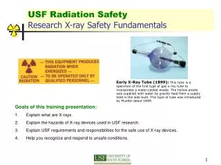
Introduction This slide show will present required safety information related to the use of Analytical X-raydiffraction (XRD) and X-rayfluorescence (XRF) instrumentation at William & Mary. The required training process is described briefly below: General awareness (contents of this slideshow) Short online quiz (link should be provided at end of slideshow) Machine specific training required before use and administered by instrument administrator All training steps must be completed and recorded prior to any unsupervised use. Analytical X-raymachines at William & Mary are primarily used for material and structural characterization. Medical use of X-raymachines requires additional, specialized training and is not addressed here. Analytical X-raysystems are defined here as a collection of components utilizing X-rayor gamma radiation to determine the elemental composition (i.e. X-rayFluorescence) or to probe the microstructure of materials (i.e. X-rayDiffraction). “Restricted Users” are those who have completed the above training and have permission to use equipment without supervision.
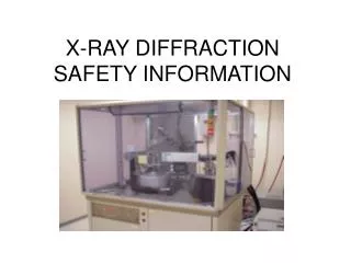
PANalytical Empyrean X-rayDiffractometer • 4-axis eulerian cradle, ideal for microstructure characterization of thin films or powdered samples. • Bragg-Brentano, grazing incidence XRD, X-rayreflectometry • data collection and analysis software including XPert Highscore • Cu K-alpha source.
Bruker Tracer III-SD X-ray Fluorescence (XRF) • Bruker Tracer III-SD XRF • Contains a Rhodium (Rh) Target • This handheld XRF allows for X-ray diffraction and elemental analysis of samples with greater flexibility over bench-top models while using the same vacuum technology. The XRF also utilizes lap-top based analytical software with live spectral displays and complete peak identification. • More information from Bruker is available at www.bruker.com.
What are X-rays? • X-rays are electromagnetic radiation which is also a form of ionizing radiation. Unlike visible light, a single photon of high-energy (short wavelength) X-rays or gamma ray radiation is capable of ionizing matter. • X-rays are an important tool in many areas of scientific research, such as the microstructure characterization or chemical identification of materials; however, there are associated risks owing to the risk of exposure to ionizing radiation. • X-rays are capable of traversing great distances and are able to penetrate many materials. There are, however, certain dense materials which block or attenuate X-rays like lead and concrete. • Most commercial instruments for analytical use (i.e. cabinet housed X-raydevices) are very well shielded and pose minimal risk of exposure to the userwhile in operation. • Owing to the health risks associated with ionizing radiation, safety training is required for the operation of such instruments that produce X-rays.
Biological effects of Radiation Exposure Radiation exposure is a well-investigated hazardous agent. Radiation levels can be readily controlled and monitored so that work may continue at a level of acceptable risk that is far less than some other common workplace activities.
The units used to measure exposure to radiation When people are exposed to radiation, the energy of the radiation is deposited in the body. As this absorption takes place, the tissue of your body may be damaged by the penetration and conversion of the radiation energy. Since absorption of radiation can damage tissue, a way to measure that damage and ensure that it is kept to a minimum is necessary. Absorbed Dose — the amount of energy of ionizing radiation absorbed per unit mass by a body, often measured in rad or Gray (Gy). Effective Dose — The tissue-weighted sum of the equivalent doses in all specified tissues and organs of the human body and represents the stochastic health risk to the whole body, which is the probability of cancer induction and genetic effects, of low levels of ionizing radiation. Dose equivalent measured in remor Sievert (Sv).
The units used to measure exposure to radiation (continued) 1 rem = 100 Sv Roughly 1 rem is the average dose received in three years of exposure to natural (e.g. background) radiation. One sievert is at the lower end of a range of doses that are likely to cause radiation sickness. 1 Sv = 0.01 rem = 10 mrem The amount of energy absorbed by tissue is a “dose”. The rate of exposure is measured by a survey meter calibrated in one (1) milliRoentgen per hour (mR/h) which is approximately 10,000 nanoSieverts per hour (nSv/h). For isotopes and X-ray energies used at W&M, 1 rad = 1 rem (approximately). 1Sv = 0.01 rem = 10 mrem is at the lower end of acute-dose radiation sickness. To get an acute-dose of 10 mrem, one needs to be exposed for one hour to a source registering 10 mR/h on a survey meter.
Background Radiation • Background radiation is radiation that can be found all around us, in the ground, rocks, air, water and our bodies. Sources of background radiation may be naturally occurring or man-made. • Natural sources of background radiation include: • Radon gas • Internal exposure from naturally occurring radionuclides in our bodies • Cosmic rays • Manmade sources of background radiation include: • Medical uses, chest X-rays, CT scans, etc. • Non-medical sources including those from industrial, occupational, or consumer sources
Average U.S. Background Radiation Sources Average annual background dose from all sources ~620 mrem
Radiation Exposure : Acute vs. Chronic • Acute Exposures— One-time event— High level doses involved (>100 rem)— Symptoms appear rapidly (within days or weeks)— Symptoms include radiation burns usual result of exposure to direct or primary X-raybeam. Hands, fingers and eyes are most commonly at risk. High-energy X-rays readily penetrate the outer layer of scan and can damage nerve endings, thus you may not feel the effects until the damage is already done. There are long term effects associated with acute exposure as well including increase rates of leukemia and other cancers. • Chronic Exposures— exposure over a long period of time— low level doses involved— Effects will appear slowly because the body has time to heal itself after exposure. Thus effects, if any, will manifest on a longer time scale (~20-30 years).— Long term effects include higher instances of cancer and cataract formation.
ALARA (As Low As Reasonably Achievable) ALARA Principle: Maintaining occupational exposures to radiation and radioactive materials As Low As Reasonably Achievable (ALARA). The risk of adverse health effects (e.g. cancer) can be minimized by minimizing your occupational radiation dose. ALARA is required by law. The guiding principles of ALARA are summarized below: 1. Time — Reduce the amount of time of exposure near a source of radiation. Plan to work efficiently near sources. 2. Distance — Maximize the distance of human tissues away from radiation sources as intensity drops with distance from the source. 3. Shielding — Ensure that there is adequate shielding between you and any radiation sources. 4. Dosimetry— Use radiation monitors where required. Always follow ALARA principles!!!
X-rayUnit Safety • The safety features of X-rayunits will vary depending on the type of unit employed (i.e. cabinet, closed beam, open beam, XRF) • Open-beam units have the highest potential for dangerous exposures to occur because they allow the user to have direct access to the primary beam while in operation. • All enclosed X-rayunits include the necessary safety features needed to lockout access to the primary beam while operating, minimizing user exposure greatly. • Portable XRF’s require strict adherence to special safety practices and procedures to avoid exposure to the primary beam. • Always familiarize yourself with your unit’s distinct safety features and design risks prior to use. • In-person, machine specific training by the machine administrator is required prior to operating any X-raydevice. • Learn and follow all standard operating procedures for each X-rayunit you operate.
XRD Cabinet Units An analytical X-raysystem is closed-beam if all possible X-raypaths (primary, scatter and diffracted) are completely enclosed so that no part of the human body can be exposed to the beam during normal operation (like the Empyrean X-raydiffractometer). These units are the safest because they prevent exposure to the primary beam by including numerous safety interlocks. While doses outside the X-raycabinet are very low, radiation exposure inside an -X-raymachine from the primary beam is very high and considered an immediate health risk if exposed. Never attempt to bypass any safety interlocks.
Portable X-ray Fluorescence Analyzers (XRF) A handheld XRF is operated in an open-beam configuration, however the sample must be in contact with the XRF before the XRF will irradiate the sample. Suitable XRF devices must have a built-in proximity sensor and backscatter detector. Both of these safety devices are used to prevent the accidental firing of the X-rays without a sample placed against the beam port.
XRF Specific Practices • Handheld XRFs deserve extra considerations due to the lack of safety features such as unit enclosures, beam shutters, beam stops and interlocks. The absence of these features allows the potential for individuals to become exposed to the primary beam and scattered radiation • DO NOT change/manipulate samples while the X-rayunit is energized. • NEVER point an XRF at an individual • ALWAYS enable safety interlocks (backscatter detector, proximity detector) • KNOW the specific provisions for XRF use as well as manufacturer’s safety procedures.
XRF Specific Practices (cont.) • BE AWARE of the primary beam’s direction. Some XRF units are emitted at an angle. • AVOIDplacing any part of your body (especially eyes or hands) near the examination area during measurement. If the sample is located on a table or bench top, make sure no feet or other body parts are beneath the working area. • If the primary beam is directed towards a wall or floor, make sure the other side of the barrier is unoccupied. • X-rays are emitted at a 53° incident angle from the examination window to the users’ left (i.e. the highest concentration of X-rays).
X-ray Exposure, First Aid and Reporting If an individual suspects that he or she has received an overexposure of external radiation from any source, he/she should immediately seek medical services and inform the Radiation Safety Officer (RSO). If you experience a radiation burn or radiation sickness, seek immediate medical attention. The exposed worker should be removed from areas in which he/she might receive more radiation and should not be allowed to return to work in such areas until authorized by the RSO following medical evaluation. Radiation Safety Officer (RSO): Eric Bradley (757) 221-2220 elbrad@wm.edu Environmental Health & Safety Office: (757) 221-2288 In any emergency, regardless of your location, dial 911. All area police (including campus police) and rescue departments use the 911 system for emergencies.
Thank you for your attention! Please proceed to the X-ray Safety Training Quiz using the link on the webpage.