Download
1 / 51
510 likes | 672 Vues
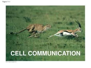
Figure 11.1. CELL COMMUNICATION. 1. 2. 3. factor. Receptor. Figure 11.2. Exchange of mating factors. a. . a factor. Yeast cell, mating type a. Yeast cell, mating type . Mating. a. . New a/ cell. a/ . 1. 2. 3. Figure 11.3. Individual rod-shaped cells.

E N D
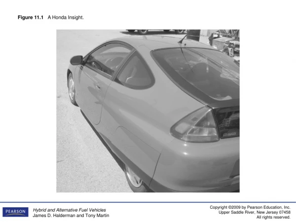
Figure 11.1 CELL COMMUNICATION
1 2 3 factor Receptor Figure 11.2 Exchange of mating factors a a factor Yeast cell, mating type a Yeast cell, mating type Mating a New a/ cell a/
1 2 3 Figure 11.3 Individualrod-shapedcells Aggregation in progress 0.5 mm Spore-formingstructure(fruiting body) 2.5 mm Fruiting bodies
Plasma membranes Figure 11.4 Gap junctionsbetween animal cells Plasmodesmatabetween plant cells (a) Cell junctions (b) Cell-cell recognition
Figure 11.5a Local signaling Electrical signalalong nerve celltriggers release ofneurotransmitter. Target cell Neurotransmitter diffuses across synapse. Secretingcell Secretoryvesicle Local regulatordiffuses throughextracellular fluid. Target cellis stimulated. (b) Synaptic signaling (a) Paracrine signaling
Long-distance signaling Figure 11.5b Endocrine cell Bloodvessel Hormone travelsin bloodstream. Target cellspecificallybinds hormone. (c) Endocrine (hormonal) signaling
1 Figure 11.6-1 EXTRACELLULARFLUID CYTOPLASM Plasma membrane Reception Receptor Signalingmolecule
2 1 Figure 11.6-2 EXTRACELLULARFLUID CYTOPLASM Plasma membrane Reception Transduction Receptor Relay molecules in a signal transductionpathway Signalingmolecule
3 2 1 Figure 11.6-3 EXTRACELLULARFLUID CYTOPLASM Plasma membrane Response Reception Transduction Receptor Activationof cellularresponse Relay molecules in a signal transductionpathway Signalingmolecule
1 2 4 3 Figure 11.7b G protein-coupledreceptor Plasmamembrane Activatedreceptor Signalingmolecule Inactiveenzyme GTP GDP GDP CYTOPLASM Enzyme G protein(inactive) GTP GDP Activatedenzyme GTP GDP P i Cellular response
2 1 3 4 Signalingmolecule (ligand) Ligand-binding site Figure 11.7c Signalingmolecule helix in themembrane Tyr Tyr Tyr Tyr Tyr Tyrosines Tyr Tyr Tyr Tyr Tyr Tyr Tyr Tyr Tyr Tyr Tyr Tyr Tyr CYTOPLASM Receptor tyrosinekinase proteins(inactive monomers) Dimer Activated relayproteins Cellularresponse 1 P Tyr P Tyr P Tyr Tyr Tyr Tyr P Tyr P Tyr P P Tyr Tyr Tyr Tyr P Cellularresponse 2 Tyr P Tyr P Tyr Tyr P Tyr Tyr P 6 ADP 6 ATP Fully activatedreceptor tyrosinekinase(phosphorylateddimer) Activated tyrosinekinase regions(unphosphorylateddimer) Inactiverelay proteins
2 1 3 Figure 11.7d Gate closed Gate closed Ions Gate open Signalingmolecule (ligand) Plasmamembrane Ligand-gatedion channel receptor Cellularresponse
EXTRACELLULARFLUID Hormone(testosterone) Figure 11.9-1 Plasmamembrane Receptorprotein DNA NUCLEUS CYTOPLASM
EXTRACELLULARFLUID Hormone(testosterone) Figure 11.9-2 Plasmamembrane Receptorprotein Hormone-receptorcomplex DNA NUCLEUS CYTOPLASM
EXTRACELLULARFLUID Hormone(testosterone) Figure 11.9-3 Plasmamembrane Receptorprotein Hormone-receptorcomplex DNA NUCLEUS CYTOPLASM
EXTRACELLULARFLUID Hormone(testosterone) Figure 11.9-4 Plasmamembrane Receptorprotein Hormone-receptorcomplex DNA mRNA NUCLEUS CYTOPLASM
EXTRACELLULARFLUID Hormone(testosterone) Figure 11.9-5 Plasmamembrane Receptorprotein Hormone-receptorcomplex DNA mRNA NUCLEUS New protein CYTOPLASM
Signaling molecule Figure 11.10 Receptor Activated relaymolecule Inactiveprotein kinase1 Activeprotein kinase1 Inactiveprotein kinase2 ATP Phosphorylation cascade ADP P Activeprotein kinase2 PP P i Inactiveprotein kinase3 ATP ADP P Activeprotein kinase3 PP P i Inactiveprotein ATP P ADP Activeprotein Cellularresponse PP P i
First messenger(signaling moleculesuch as epinephrine) Figure 11.12 Adenylylcyclase G protein GTP G protein-coupledreceptor ATP Second messenger cAMP Proteinkinase A Cellular responses
EXTRA-CELLULARFLUID Signaling molecule(first messenger) Figure 11.14-1 G protein DAG GTP G protein-coupledreceptor PIP2 Phospholipase C IP3 (second messenger) IP3-gatedcalcium channel Endoplasmicreticulum (ER) Ca2 CYTOSOL
EXTRA-CELLULARFLUID Signaling molecule(first messenger) Figure 11.14-2 G protein DAG GTP G protein-coupledreceptor PIP2 Phospholipase C IP3 (second messenger) IP3-gatedcalcium channel Endoplasmicreticulum (ER) Ca2 Ca2(secondmessenger) CYTOSOL
EXTRA-CELLULARFLUID Signaling molecule(first messenger) Figure 11.14-3 G protein DAG GTP G protein-coupledreceptor PIP2 Phospholipase C IP3 (second messenger) IP3-gatedcalcium channel Variousproteinsactivated Cellularresponses Endoplasmicreticulum (ER) Ca2 Ca2(secondmessenger) CYTOSOL
Growth factor Reception Receptor Figure 11.15 Phosphorylationcascade Transduction CYTOPLASM Inactivetranscriptionfactor Activetranscriptionfactor Response P DNA Gene NUCLEUS mRNA
Figure 11.18 Signalingmolecule Receptor Relay molecules Activationor inhibition Response 2 Response 3 Response 5 Response 4 Response 1 Cell B. Pathway branches,leading to two responses. Cell D. Different receptorleads to a differentresponse. Cell C. Cross-talk occursbetween two pathways. Cell A. Pathway leadsto a single response.
Ced-9protein (active)inhibits Ced-4activity Figure 11.21a Mitochondrion Ced-3 Ced-4 Receptorfor death-signalingmolecule Inactive proteins (a) No death signal
Ced-9(inactive) Cellformsblebs Figure 11.21b Death-signalingmolecule ActiveCed-4 ActiveCed-3 Otherproteases Nucleases Activationcascade (b) Death signal
Figure 11.22 Cells undergoingapoptosis Space betweendigits 1 mm Interdigital tissue
Chapter 12 The Cell Cycle
100 m (a) Reproduction Figure 12.2 200 m (b) Growth and development 20 m (c) Tissue renewal
Figure 12.4 Sisterchromatids Centromere 0.5 m
ChromosomalDNA molecules Chromosomes Figure 12.5-3 Centromere 1 Chromosomearm Chromosome duplication(including DNA replication)and condensation 2 Sisterchromatids Separation of sisterchromatids intotwo chromosomes 3
INTERPHASE Figure 12.6 S(DNA synthesis) G1 Cytokinesis G2 Mitosis MITOTIC(M) PHASE
Figure 12.7a Prometaphase G2 of Interphase Prophase Fragments of nuclearenvelope Centrosomes(with centriole pairs) Early mitoticspindle Nonkinetochoremicrotubules Chromatin(duplicated) Aster Centromere Plasmamembrane Kinetochore Nucleolus Kinetochoremicrotubule Chromosome, consistingof two sister chromatids Nuclearenvelope
Centrosome Figure 12.8 Aster Metaphaseplate(imaginary) Sisterchromatids Microtubules Chromosomes Kineto-chores Centrosome 1 m Overlappingnonkinetochoremicrotubules Kinetochoremicrotubules 0.5 m
Figure 12.9b CONCLUSION Chromosomemovement Kinetochore Microtubule Tubulinsubunits Motor protein Chromosome
Figure 12.7b Metaphase Anaphase Telophase and Cytokinesis Nucleolusforming Metaphase plate Cleavagefurrow Nuclearenvelopeforming Spindle Centrosome atone spindle pole Daughterchromosomes
Figure 12.11 Chromatincondensing Nucleus 10 m Nucleolus Chromosomes Cell plate 3 4 5 2 Anaphase 1 Prophase Metaphase Telophase Prometaphase
Figure 12.11a Chromatincondensing Nucleus Nucleolus 10 m 1 Prophase
Figure 12.11b Chromosomes 10 m 2 Prometaphase
Figure 12.11c 10 m 3 Metaphase
Figure 12.11d 10 m 4 Anaphase
Figure 12.11e 10 m Cell plate 5 Telophase
Figure 12.10 (a) Cleavage of an animal cell (SEM) (b) Cell plate formation in a plant cell (TEM) 100 m Vesiclesformingcell plate Cleavage furrow Wall of parent cell 1 m New cell wall Cell plate Daughter cells Contractile ring ofmicrofilaments Daughter cells
Cell wall Origin ofreplication Plasma membrane Figure 12.12-4 E. coli cell Bacterial chromosome 1 Chromosomereplicationbegins. Two copies of origin 2 Origin Origin Replicationcontinues. 3 Replicationfinishes. 4 Two daughtercells result.
Diatoms andsome yeasts (c) Bacterialchromosome (a) Bacteria Figure 12.13 Chromosomes Microtubules (b) Dinoflagellates Intact nuclearenvelope Kinetochoremicrotubule Intact nuclearenvelope Kinetochoremicrotubule (d) Most eukaryotes Fragments ofnuclear envelope
G1 checkpoint Figure 12.15 Controlsystem S G1 G2 M M checkpoint G2 checkpoint
Figure 12.16 G0 G1 checkpoint G1 G1 (a) Cell receives a go-ahead signal. (b) Cell does not receive a go-ahead signal.
Figure 12.17a M M G1 G2 G1 G2 M S G1 S MPF activity Cyclinconcentration Time (a) Fluctuation of MPF activity and cyclin concentration during the cell cycle
Figure 12.17b G1 S Cdk Cyclin accumulation M G2 Degradedcyclin G2checkpoint Cdk Cyclin isdegraded Cyclin MPF (b) Molecular mechanisms that help regulate the cell cycle