Download

1 / 96
980 likes | 1.06k Vues
Explore the structure, function, and development of the skin, the body's heaviest organ, covering topics from epidermis and dermis to skin lines and types.

E N D
David Kachlík Skin and skin organs Integumentum commune
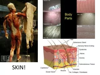
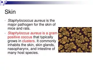
Skin = Cutis (Gr.derma) • heaviest organ in the body • 16% weight, 1,2-2,3m2 • cuticle (epidermis) • cutis (corium,dermis) hypodermis (tela subcutanea; subcutis,hypodermis) • does not belong to skin as an organ • skin derivatives
Skin - function • protection • termoregulation • Sudorific glands • Changes in the blood flow • (respiration) – perspiratio insensibilis • excretion • Absorption of medicaments • immunity • metabolism (ergosterol → vitamin D) • Emotions and psychic
Skin - relief • skin grooves(sulci cutis) – in between of them rhomboid skin fields • Tactile crests (cristae cutis) – 9 types according to Purkynje → dactyloscopy • Tactile pillows (toruli tactiles) – 10 on hand (ce thenar) • Bending sulci (lineae distractiones) • Skin vincula (retinacula cutis) • (retinaculum caudale) • striae (striae cutaneae) – growth, gravidity, obesity
Bending sulci (lineae distractiones) • Sulci at joints, wrinkles • sulcus mentolabialis, nasolabialis, suprapalpebralis, infrapalpebralis • sulcus gluteus, crena ani • hand – Purkynje - chiromantia • linea oppositionis pollicis (vitalis) • linea manus clausae (cephalica, naturalis) • linea occlusionis dig. trium ulnarium (mensalis) • sulcus cutaneus intercarpalis (linea rasceta) – most proximal carpal • linea restricta – middle carpal
Lines of fissility podle Kraisla = Langers skin lines; cleavage lines; tension lines Runs in direction of fibril bundles in dermis (stratum reticulare) Vertical to direction of strongest pull Correspond to wrinkles on surface of skin Important in plastic surgery and for surgery incisions "lines of greatest tension" (Kraissls lines) – defined on living – vertical to muscle position orientation Borges lines – according to lines present in feebled skin Kraissl + Borges lines better effect
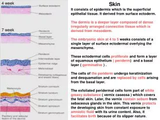
Skin - development ectoderm → epidermis (surface) mesoderm → dermis → Langerhans cells Neural crest → melanocytes → Merkels cells
Epidermis - development during 1st and 2nd trimester • primordium – 1st layer of epidermal cells • proliferation of surface ectoderm → • PERIDERM • BAZAL GERMINAL LAYER (= prospective stratum basale) • Formation of epidermal crests (dermatoglyfs) • Migration of cells from neural crest – melanoblasts + Merkels cells • Keratinization and deskvamation = VERNIX CASEOSA of newborn • Protects from exposition to amniotic fluid • Facilitate delivery
Dermis - development • From somatopleura of lateral mesoderm • Part from dermatomes • in 11th week starts production of fibers • COLLAGEN • ELASTICS • Continually happening capillarization and innervation • epidermis and dermis mutually invaginate
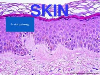
Types of skin • Thick type (smooth and glabrous skin) • Palms and soles • 400-800 μm thick epidermis • Thin type (hairy skin) • 75-150 μm thick epidermis • Missing stratum lucidum • Overall thickness depends on topography • back – 4 mm • Hairy part of head – 1,5 mm
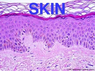
Epidermis • keratinocytes • Squamous epithelium – 5(6) layers • stratum basale • stratum spinosum • stratum granulosum • stratum lucidum • stratum corneum • stratum disjunctum • melanocytes • Langerhans cells • Merkel cells
Epidermis - Stratum basale • 1 layer of cylindrical / cuboid elements • bazophilic • Desmozomes, hemidesmosomes • mitotic activity • Whole epidermis recreates within 15-30 days • psoriasis – fastened to 7 days • 10 nm intermediary filaments (cytokeratins) • Towards surface more numerous • In thick type of skin are Merkel cells
Epidermis - Stratum spinosum • Spiked cells, on summits are desmozomes • cytokeratins (tonofilaments) converge into desmozomes • Multiplied on palms and soles • Numerous mitoses • Together with stratum basale forms STRATUM GERMINATIVUM MALPIGHI • Langerhans cells
Epidermis - Stratum granulosum • 3-5 layers of cells • Polygonal with centrally located nucleus • 2 types of granules: • BAZOFILIC granules = keratohyalin • LAMELAR granules = Odlandcorpuscles (content excreted into intercellular space serves as cement against pass of foreign bodies)
Epidermis - Stratum lucidum • thin • Flat eosinophilic cells • Nucleus and organels not well visible • Mostly filaments in electrondense matrix • Visible desmozomes • ELEIDIN (very structurated)
Epidermis - Stratum corneum • 15-20 layers of flat cells = cells of the corneal layer • Nuclei and organels not present • Effect of lyzosomal enzymes • cytoplazm filled by KERATIN • Composed scleroprotein cemented by basic substance from keratohyalin granules • stratum disjunctum – releasing cells
Coloring of skin • melanin (melanocytes) • karoten • Number of vessels in dermis • Color of blood in vessels of dermis
Melanocytes • In between elements of stratum basale, in hairy follicles • Round bodies with extensions into epidermis • create melanin (eumelanin) • Dark brown pigment • In foxy hairs feomelanin • Synthesis o tyrosinase and deposition in sacs (melanosomes) injected into keratinocytes (melanofors)
Melanin production • Tyrosinase converts • tyrosine into 3,4 -dihydroxyfenylalanin (DOPA) • DOPA into dopachinon • dopachinon converts into melanin • microscopically 4 stages (events in melanosome) I. Bland grained material, tyrosinase located on protein matrix (= premelanosoma) II. Parallel filaments - melanin on protein matrix with period 10 nm ( = vesicula striata) III. period ceases with higher amount of melanin IV. sac fully filled with melanin, visible LM, lacks ultrastructure
Melanocytes • produce melanin • Mature melanin granules = melanosomes • cytokrine secretion of melanosomes into keratinocytes
Secretion of melanin • cytokrine secretion via extensions • Concentration in supranuclear region • Protection of mitotic cells from UV • epidermal melanin unit • 2 phases of skin darkening • Darkening of existing melanin • Production of new sacs • albinismus – inborn defect of melanin production (total) • vitiligo – degeneration and dissapearance of melanocytes (local) • chloasma uterinum – irregular spots, most common in gravidity (head, forearm) • chloasma/melasma suprarenale: Addison disease–increased pigmentation
Other cells • Langerhans cells (Dendrocyti) • macrophages • 2-8 % cells of skin • Mostly in stratum spinosum • Merkelovy buňky (Epithelocyti tactilis) • In thick type of skin - stratum basale • Dark granules of uncertain composition • Based on free nerve endings • mechanoreceptors - Complexus epithelialis tactus • Derivatives of neural crest !
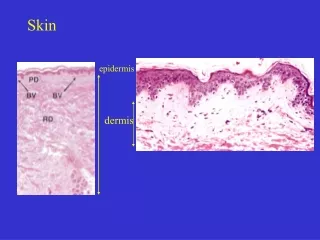
Dermis - composition • Fibrous layer under basal membrane of epidermis • thickness up to 4 mm (back) • extensions - dermal papillas • Correlates with epidermal pegs • 2 slurry bordered layers • stratum papillare and reticulare • Rich capillary bedstream • Presence of skin derivatives
Dermis - Stratum papillare • Forms great part of papills • Thin tissue • Common fibrous components • ANCHORING FIBERS inserting into basal membrane
Dermis - Stratum reticulare • Thick tissue • Plenty of fibers especially collagen type I • dermatan-sulfate • elastic mesh inserting into basal membrane • ageing • Ehlers-Danlos illness, cutis laxa (defect production of collagen fibers)
Dermis - function • Reinforcement of epidermis • pegs = papillas • Anchoring structures • Blood supply • Termoregulation and regulation of blood pressure • Enabeling of inputs • Free nerve endings (terminationes neurales liberae) and other sensory bodies
Skin - supply • arteries – deep and superficial net • Always constant stalk for particular area skin folds for replantations • capillaries - a-v anastomoses • veins – the same • lymph – capillaries subcapillary mesh collectors • nerves – missing parasympaticus ! • Nerve endings (free x corpuscles) • 5th sense = touch – somatosensory fibers • sympatic postganglionic vasomotoric fibers – adrenergic – visceromotor fibers • sympatic fibers for sudoriferous glands – cholinergic !
Skin diseases I. Pemphigus vulgaris • Autoimmune illness • Circulating IgG anti bodies against surface antigens of cuboid epithelium associated with desmozomes • Binding of antibody causes activation of proteases, breakdown of epidermis and formation of blisters • Intraepidermal blisters • Located suprabazally
Skin diseases II. Bulous pemfigoid • autoimmune illness: circulating IgG anti bodies binding to antigens BP1 and BP2 in basal membrane area • Binding of anti bodies leads to complement activation, defect of tissue and blisters • Subepidermal blisters with cover formed by relatively normal epidermis
Skin diseases II. Skin tumors • 1/3 of all tumors • Originate from keratinocytes of stratum germinativum • bazalioma • spinocellular carcinoma • - are recognized early • Originate from melanocytes– melanoma • growth through BL,penetrates into vessels, metastasing • vzdálené mestastázy i z malých nádorů
Skin derivatives • hairs (pili) • lanugo /flumina, vortices/ • hairs (capilli), eyebrow (supercilia), eyelashes (cilia), beard (barba), tragi (external auditory canal), vibrissae (nose), hirci (armpit), pubes, tactile (sinusoids) • nails (ungues) • Skin glands (glandulae cutis) • Are derivatives of epidermis
Hairs (pili) • Everywhere except for palms, soles, lips, glans penis and labia minora • Allocation, density and color depends on: • Sex, age, race and body area • Effect of hormones (androgens, T3,T4, corticoids) • Growth periods (anagen) • Quiescent periods (katagen, telogen)
Hair growth • anagen - 3 years/ 1000 days • katagen - 3 weeks/ 10 days • telogen - 3 month/ 100 days Perish of bulb and its renewal 0,4 mm/day Falling out – up to 100 hairs per day – if more effluvium areas w/o hairs - alopecia Influence of: hormones, nutrition, toxic (infections, chemoterapy, autoimmune)