Download
1 / 11
120 likes | 233 Vues
Explore the fascinating history of microscopes from Leeuwenhoek to modern electron microscopes. Learn about different types of microscopes, their parts, and how to enhance specimen visibility. Discover the capabilities and limitations of electron microscopes.

E N D
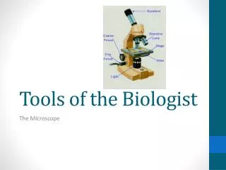
Tools of the Biologist The Microscope
History • Antonie von Leeuwenhoek—Born in Holland in 1632 • First to observe living bacteria & drew them. • Also looked at sperm, protists, blood • 1stsimple scope • Made over 500 "microscopes"
History • Robert Hooke (1665) – Used compound scope to examine cork. • Coined the term “cell” referring to the many little boxes. • Actually saw dead plant cells.
Types of Microscopes • Simple microscope – Hand lens(magnifying glass) • Only has 1 lens • 3-40 times magnification
Types of Microscopes • Compound Light Microscope – The type we use in our labs • Has two sets of lenses – Ocular (eye piece) & Objective (near the object being viewed) • Most commonly used microscope • Uses lenses and light to magnify & view the specimen • Total magnification on our scopes = 40 – 1000 times • Total Magnification = Ocular (10X) x Objective (40X)
Microscope Parts Ocular – eye piece. Magnifies 10x Neck – Supports the eyepiece. Objectives – 4 – 100x magnification Arm – Supports neck and objectives. Carry by this Stage and clips – Holds slide in place Adjustments – Coarse & Fine. Focuses image Diaphragm – Controls the amount of lightcoming through the stage Light – Electric light source Base – Bottom of scope. One hand goes underneath
Making specimens visible • Since most of the specimens we observe will be clear to near clear, what could be done to enhance the image we view through the scope? • Adjust the diaphragm to allow less light to come through • Use a stain to make transparent specimens visible. Ie. Iodine, methyl blue • Specimens must be sliced very thin. Blood cells Morel spores Endospores Colon x section Phytoplankton
Electron Microscopes • Uses electromagnets and streams of electrons to view a specimen • Limit of Resolution is 1000x finer than light microscope • 200,000 – 1,000,000 x magnification • Two types
Transmission EM • Transmission Electron Microscope (TEM) 1931 (Germany) • Image is seen on a fluorescent screen • Specimen must be thinly slices and coated in carbon. • Gives a 2D of specimen • Specimen must be dead Staphylococcus Herpes simplex virus E. Coli aureus
Scanning EM • Scanning Electron Microscope (SEM) – 1935 (Germany) • Gives a 3D image and coated in Au or Ag • Electrons scan around specimen • Shows only the outside of the specimen • Gives very clear details of surface structures Weevil Tick DiatomRadiolarian
Limits of the Electron Microscope • Specimens must be very thin • Specimens must be stained or coated • Specimens must be dried out (Mounting chamber is vacuum sealed • Specimens must be dead • BLACK AND WHITE images only!