Download
1 / 7
100 likes | 701 Vues
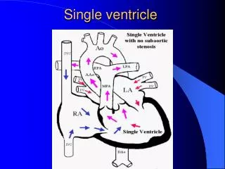
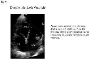
Fig 53. Double inlet Left Ventricle. LV. Apical four chamber view showing double inlet left ventricle. Note the presence of two atrioventricular valves connecting to a single morphology left ventricle. MLV. RV. RA. LA. LA. LA. RA. Fig 54. Tricuspid atresia. LV. RV. vsd. LA. RA.

E N D
Fig 53 Double inlet Left Ventricle LV Apical four chamber view showing double inlet left ventricle. Note the presence of two atrioventricular valves connecting to a single morphology left ventricle. MLV RV RA LA LA LA RA
Fig 54 Tricuspid atresia LV RV vsd LA RA Apical four chamber view from a patient with an absent right atrioventricular connection (tricuspid atresia). The position of the tricuspid valve is replaced by strand of tissue (arrow). The right ventricular cavity is very small. A perimembranous VSD is also present.
Fig 55 CLASSICAL GLENN BIDIRECTIONAL GLENN FONTAN SVC SVC RPA LPA PA PA RA RA RA IVC IVC TOTAL CAVOPULMONARY CONNECTION SVC PA RA IVC
Fig 56 Tricuspid atresia Apical four chamber view of a univentricular atrioventricular connection with atresia of the right sided atriovenricular valve (tricuspid atresia).The patient had a Fontan type repair (right atrium to pulmonary artery connection). The valve between the IVC and right atrium can just be seen in the right atrium (arrow). MLV LA RA
Fig 57 TCPC Apical four chamber view of a patient with a univentricular atriovenricular connection and a common atrium, post TCPC. Note the TCPC pathway (arrow) at the back of the atrium. V Common atrium
Fig 58 Tricuspid atresia post Fontan operation Apical view of a univentricular atrioventricular connection resulting from atresia of the right-sided atrioventricular valve (tricuspid atresia). The patient had a Fontan type operation. Note that the right atrium is severely dilated with thrombus inside. LV LA RA
Fig 59 Fontan: The giant RA and large clot RA RA TOE image taken from a patient with a double inlet left ventricle post Fontan (atria-pulmonary connection) repair. Note the severely dilated right atrium with giant thrombus and spontaneous contrast inside the atrial chamber (arrow).