Download
1 / 64
640 likes | 793 Vues
Patophysiology of blood and lymph circulation in lower extremities Assoc. prof . Jana Plevkova MD, PhD 20 12 Department of Pathophysiology Jessenius Faculty of Medicine in Martin.

E N D
Patophysiology of blood and lymph circulation in lower extremities Assoc. prof. Jana Plevkova MD, PhD2012Department of PathophysiologyJessenius Faculty of Medicine in Martin
Blood circulation is sophisticated system which conduct the blood from the heart and lungs into the tissues- it's main function is to provide suitable metabolic supply to the cells /oxygen, substrates/, aw well as the cleavage of metabolic products from the tissue • vessels – resistance, capacity, capillary network, other specific vascular structures • high pressure / low pressure part of circulation • regional specific circulations
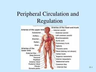
Arterialsystemoflowerextremities source :http://education.yahoo.com/reference/gray/subjects/subject/157
Principles of blood flow science discipline analyzing the flow of the blood within the circulation P. .r4 8..l Q Q – blood flow P – pressure gradient between two different points of the tube /vessel/ r – vessel diameter l – vessel length - blood viscosity
Arterial system of lower extremities -a part of high pressure circulation Energy of the heart ejection /systolic effort/ continues into 1)frontal pressure, responsible for the forward movement of the blood within the vessel 2)lateral pressure affecting the wall of the artery causing it's distension, giving the base for the pulse wave laminar flow(flow within individual layers smoothly gliding one on another ) - This mutual gliding is responsible for the tangential tension among the layers and most external layer and endothelium, as well (shear stress)
shear stress – this mechanism is able to influence some of the regulatory and secretory functions of the endothelium Turbulent flow within the blood stream could be physiological only at the sites of wide vessels • aortal arc, pulmonary artery, site of the vessel branching, sudden bending of the vessel Predisposition for turbulent blood flow -wide vessel -sudden change of the diameter - high blood flow velocity - low blood viscosity -uneven endothelial surface
The blood flow in the arteries has three phases, due to elastic properties of the vessel wall 1st phase – forward movement of the blood caused by ventricular contraction – systolic ejection. Vessel wall is distended during this phase, because of lateral pressure applied onto the vessel wall 2nd phase is characterized by return of the distended vessel diameter into the former size – elastic recoil the flow is directed to the beds with low resistance 3rd phase of the diastolic period – the flow is directed forward again
Regulation of the blood flow in lower extremities Relative constant pressure gradient – diving pressure is constant, so regulation is secured predominantly by the change of vessel resistance – change of vessel diameter • myogenic (Bayliss regulation) • metabolic (autoregulation) lactic acid, CO2, H+, K+, adenosin • other humoral factors (catecholamine, histamine, acetylcholine, angiotensin) • nerve regulation - sympathetic fibers
Endothelium – is not only a mechanical barrier It has high metabolic activity, participates in vessel reactivity, regulation of thrombogenesis, influences the functions of circulating cells Endothelial surface – 500 - 1000 m2 – contact surface for the mediation of „signals“ between circulating cells and intimal surface It seems to be the largest endocrine organ /1500g/ Metabolic and secretoric systems influence mainly vessel tone therefore resistance and blood flow Physiological tendency to vasodilatation
Endothelial vasodilators • production of NO – from L arginine by NO synthasis – enzymatic process, new molecule of NO is released into the smooth muscle cells layer beneath the endothelium – activation of guanylatcyclase - production of cGMP leads to relaxation of muscle cells and thus to vasodilatation • production of NO is responsible for permanent „natural“ tendency to vasodilatation in arterial system • production of NO is stimulated by – shear stress, platelets derived molecules like (ATP, ADP, serotonin), vessel distention – flow dependent dilatation • NO is dominant vasodilator in basal conditions • endothelium produce also other vasoactive substances like - PGI2 (prostacyclin) PGE2, PGD2
Endothelial vasoconstrictors • endothelins, tromboxan A2, nonstable endoperoxides, and molecules of local RAA system • Endothelins (1, 2, 3) – group of peptides containing 21 AA, derived from molecule of proendothelin, which is fragmented to active molecules • ETA a ETB receptors – vasoconstricting response, long lasting effects involve proliferative effects on smooth muscle cells within the vessel wall
Endothelial dysfunction Functional changes of the endothelial cells • predominance for production of vasoconstricting mediators • increased production of cytokines • increased permeability for plasmatic proteins and lipoproteins • predominance of procoagulating processes • increased production of CAM molecules
Pathogenesis of diseases of arterial system of lower extremities Diseases with different ethiology may have the same, or very similar signs and symptoms • So it is not correct to talk about „arterial occlusion“ only Ischemia of lower extremities • acute – sudden onset of ischemic attac /dangerous, bc of the risk of the lost of the extremity/ • chronic – long lasting ischemization, trophic changes... - Degenerative processes - atherosclerosis /ATS/ - Aneurysmal arterial disease - Inflammation and thrombosis - Vasospastic disease
Atherosclerosis obliterans Risk factors fatty and cholesterol containing diet less fruits/vegetable/fiber diet hypercholesterolaemia smoking hypertension low concetration of HDL + family history for CVS diseases obesity increased fibrinogene level male sex
(Atherosclerosis obliterans - ASO) patogenesis of ATS – response of the vessel wall to injury • functional changes of endothelial cells • deposition of lipid particles into the vessel wall with subsequent reaction – creation of fibromuscular plaque • chemotactic activity of monocytes – fagocythosis of lipid particles of macrophages (foam cells) lipid plaque Formation of ATS plaques of different size and position in the arterial system of lower extremities
Consequences • turbulent flow at the site of the plaque location • presence of the plaque may lead to „serious“ stenosis limiting the blood flow • damage of the vessel wall due to plaque may lead to weakening of media and formation of aneurysma • bleeding into the plaque with possibility for formation of false aneurysma • dysrupture of the plaque with subsequent thrombosis of the artery • abruption of the plaque and embolization of those fragments into more peripheral circulation acute or chronic ischemization of extremity
Arterialaneurysms http://www.daviddarling.info/encyclopedia/A/aneurysm.html http://images.rheumatology.org/viewphoto.php?imageId=2861575&albumId=75674
Arterial aneurysms – localized dilatation of the vessel wall • True aneurysms – consist of all three layers of arterial wall, usually has fusiform or circumferential shape, the underlying condition for such a dilatation is weakening of the vessel wall due to some pathological process – mainly ATS The damage of the vessel wall with weakening of media could be acquired(atherosclerosis, inflammation, toxic ifl.) or inherited (syndroms with weak connective tissue like sy. Marfan) 2)False aneurysms – extravascular accumulation of blood with disruption, two or all three vascular layers the wall of the aneurysms is formed by thrombus and adjacent tissues, or adventitia False aneurysms is usually consequence of trauma, or complication of ATS plaque
http://medical-dictionary.thefreedictionary.com/false+aneurysmhttp://medical-dictionary.thefreedictionary.com/false+aneurysm
Whatever the cause, the aneurysm becomes progressively larger ! • tension within the vessel wall is directly influenced by the diameter and the lateral blood pressure • localized dilatation of the vessel at the site of aneurysms leads to increase of diameter and therefore enhance the tension within the vessel wall, what again may enhance it´s enlargement Consequences • Ischemia below the location of aneurysma • Acute thrombosis at the site of aneurysma • Dysrupture of aneurysma • Embolization of the thrombus into the more peripheral circulation
Inflamatory diseases – Thrombangitisobliterans Burger´s disease Inflammatory disease of small peripheral arteries • chronic inflammatory process • inflammation is localized at the intima of affected vessels, and thrombosis is just a secondary consequence of it • affected vessels are prone to vasospasm • affected are mainly tibial and plantar arteries
Etiopathogenesis of the disease is unknown affected population - men 20 – 40 yrs, smokers, autoimmunity Disease has three stages 1)inflammatory and spastic phase (phlebitis saltans, migrans) 2)obliterative phase with symptoms and signs of ischemia 3)gangrene
Vasospastic diseases Attacks of sudden constrictions of small diameter arteries and arterioles of upper and also lower extremities, commonly fingers, sometimes toes Raynaud disease – primary vasospastic without any obvious or clear cause, most affected are young ladies Raynaud phenomenon – syndrome, secondary problem usually linked with systemic collagen diseases, autoimmune diseases, toxic influences, long lasting vibration exposition, ...
Raynaud disease In spite the cause is unknown, there was identified hypetrophy of myoepithelial cells, which participate in regulation of blood supply to the capillary bed and hyperplastic changes of a-v- anastomoses • cold or emotional stimulus is usually the provoking factor, leading to severe vasoconstriction, blood is redirected through a-v anastomoses into the venous system, while the capillaries are compromised Typical three phasic color changes of the skin, changes are always symetric a)sudden pallor (dogiti mortui) b)followed by cyanosis c) finally redness caused by reactive hyperaemia
Raynaud phenomenon • It is only a sign (manifestation/ of other primarily well defined disease • Example: systemic lupus erythematodes, primary pulmonary hypertension, some endocrine disorders – myxedema-, exposition to vibrations, intoxication with ergot (claviceps purpurea) Symptoms are asymetric, affected persons are both men and women
All mentioned diseases may lead to acute or chronic progressive obliteration of the vessel lumen. Obliteration of the lumen increases the vessel resistance. Increased resistance means - decreased blood supply to the affected region with a possibility of ischemia.
http://lifeinthefastlane.com/2011/01/cardiovascular-curveball-012/http://lifeinthefastlane.com/2011/01/cardiovascular-curveball-012/ http://www.bellevuepodiatrist.com/raynauds-phenomenon-red-white-and-blue-toes/
Occlusive arterial disease Chronic occlusive arterial disease ischemia as a result of arterial obstruction Obstructive arterial lesions occur more frequently in the lower extremities than in the upper extremities.Obstruction influencing the blood flow to lower extremities is usually localised. at: - aortoiliac level - femoropopliteal level - popliteo-tibial level
Development - arterial lumen is progressively narrowed resistance to blood flowblood flow to the tissue below the lesion is reduced tendency to tissue ischemia - vessel lumen must be reduced by approximately 50% in diameter or 75% in crossectional area to produce clinically significant interference with blood flow - in combination (stenosis occurring in sequence), less significant lesions can seriously impair blood flow
Stenosis less than 75% of crossectional area is not compromising blood flow during the rest condition, but during the physical exercise – it could interfere with the blood supply leading to ischemia At the site of stenosis and below the stenosis we can see changes of the blood flow like • acceleration of the blood flow at the site of stenosis • turbulent blood flow below the stenosis with recirculation of the blood, whirls (murmors present above the affected vessel • poststenotic dilatation with possibility for thrombogenesis
Example for stenosis occurring in sequence, less significant lesions can seriously impair blood flow When small nonsignificant stenosis may lead to ischemia? • During physical exercise (O2 requirements, vasodilatation in working muscle, decrease of driving pressure) • During elevation of the extremity (no hydrostatic effect supporting the blood supply)
Intensity of ischemic damage depends on – • the site /level/ of the vessel occlusion – aortoilic, femoropopliteal, popliteotibial level • extent and seriousness of stenosis • time course of occlusion development /acute vs. chronic/ • presence and quality of collateral circulation Collateral circulation is unique and important compensatory mechanism of long lasting, progressively worsening ischemia. Increased resistance of affected arteries is responsible for „opening“ of collateral circulation. Mainly muscular arterial branches could be base for collateral circulation.
Symptoms and signs of occlusive arterial disease chronic course - usually ATS • claudicatio intermittens • pain at rest • no pulse • postural changes of the skin color • temperature gradient bellow and above stanosis • neurologic symptoms – paresthesis • trophic changes of the skin, hair, nails • atrophic muscles and soft tissues • ulceration and gangrene – (dry, wet)
Symptoms and signs of occlusive arterial disease acute course – thrombosis, embolisation, trauma • dominant severe ischemic pain • no pulse • distal part below the stenosis is pale • temperature gradient • decreased filling of superficial venous system • no trophic changes – there is no time for their development
Occlusive arterial disease in patients with DM macroangiopathy – ATS(in DM patients is accelerated) • hypertension • hyperlipidaemia and dyslipidaemia • impaired nutrition of vessel wall because of dysfunction of vasa vasorum microangiopathy Damage of small diameter arteries and capillaries by generaliezed chronic complication of DM endothelial damage (diffusion of glucose into the cells, change into sorbitol, endothelial swelling – endothelial dysfunction....) In this group of patients – ischemic problems with lower extremities are very common, shifted to „younger“ age, and are usually complicated by immunodeficiency and metabolic disorder /worse healing of wounds/
http://www.surgery.usc.edu/divisions/vas/legpainandlowerextremityarterialdisease.htmlhttp://www.surgery.usc.edu/divisions/vas/legpainandlowerextremityarterialdisease.html
Venous system of lower extremities http://www.chivatechnique.com/veins.php
Blood flow in veins – lower extremities • vis a tergo – rest of left ventricle ejection energy • vis a fronte – suction of right atrium caused by up and down movement of AV junction • negative thoracic pressure during inspiration • craniocaudal movement of diaphragm • corresponding changes of abdominal pressure • horizontal body position • rhythmic compression and decompression of deep venous system by muscles /walking/ • valves • plantar venous mechanism • pulsation of commitant arteries
superficial venous system (10% of venous returs) deep venous system (90% of venous return) system of perforating veins Physiological pattern of venous return from lower extremity: Venous system of lover extremities
Function of the valves If the valves are working properly: • they completely prevent pervasion of the blood from deep into superficial system • they redirect central blood flow within the deep system • they prevent retrograde blood flow from the upper level, if once the blood was ejected by muscle pump to upper level. During the muscle relaxation they completely prevent backward blood flow to more distal levels.
Trombophlebitis Pathological process affecting mainly superficial venous system. Primary affection is inflammation of spa. vein by the process spreading from the surrounding tissue Creation of thrombus is usually secondary phenomenon superimposed to inflammation. Thrombus is fixed to the vein wall by fibrin connections between the thrombus and inflamed vessel wall. The process is usually localized to the site of inflammation, no or minimal systemic symptoms, only local signs of inflammation. No risk of emboli, b/c the thrombus if fixed onto the vein wall, and as well is localized within the superficial system.
Deep venous thrombosis DVT - intravital coagulation of the blood inside the vessels - physiological mechanisms against thrombosis • continual blood flow • intact endothelium • balance in production of pro and anti coagulating factors Impairment of those three mechanisms , known as Virchow´s trias has crucial role in pathogenesis of DVT.
Predisposing factors for DVT • history of DVT • immobilization (slowness of venous return from LE DK) • senior age(polymorbidity, dehydration, change of rheologic properties of the blood) • obesity, malignity (production of procoagulating factors) • heart failure, decompensation (venous congestion in backward failure) • surgical procedures, trauma (release of tissue tromboplastin) • pregnancy, puerperium, abortion (occlusion of pelvic veins by pregnant uterus, enhanced coagulation, tissue of trophoblast)
Pathogenesis Slowing of the blood flow during immobilization, or in case of procoagulative status the blood there is a possibility of deposition of small amounts of fibrin at the site of vein valves The fibrin deposition is growing progressively by apposition of „new“ fibrin fibers and platelets trapped into the fibrin matrix – formation of thrombus The thrombus is a serious occlusion, which impairs or completely blocks the venous return through the affected deep vein
http://www.jaapa.com/cvi-and-pad-a-review-of-venous-and-arterial-disease/article/136677/http://www.jaapa.com/cvi-and-pad-a-review-of-venous-and-arterial-disease/article/136677/
the blood flow in central direction through affected vein is blocked by the thrombus • muscle contraction eject the blood into the surrounding deep vein /deep veins are usually doubled/ via anastomosis • this deep vein provide central flow of the blood, which is of course limited – so venous return as a complex process is impaired • In this phase of DVT the blood does not flow into the spf. System, b/c perforators and their valves are not affected/destroyed, YET. Venous return from affected extremity
Muscle contraction ejects the blood into three directions • through partially recanalized vein upwards • through anastomosis into surrounding deep vein • through perforating veins /which valves are destroyed/not working / into the spf. system. • During muscle relaxation – spf. veins are emptying only partially into the deep veins, with sudden balancing of the pressure in both systems. • Blood is cumulating in spf. veins – leading to permanent hypertension in spf. system and recanalized part of deep venous system. Venous return in a stage of recanalisation
Venous return after total recanalisation Total recanalisation means healing of the vein, desobliteration, consequence of this process unfortunately is fibrotisation, or destruction of the valves. During muscle contraction the blood from deep vein is ejected into three directions -to SPF veins b/c of insufficient valves of perforating veins -to central direction through recanalised vein upwards -to central direction through surrounding deep vein Muscle relaxation: tendency for downward blood flow with rise of pressure in deep system, therefore emptying of spf. veins which had to be enhanced by the „suction“ of negative pressure is limited