Hemodynamic Disorders
Hemodynamic Disorders. Hemodynamic Disorders Thromboembolic Disease Shock. Overview. Edema (increased fluid in the ECF) Hyperemia (INCREASED flow) Congestion (INCREASED backup) Hemorrhage (extravasation) Hemostasis (keeping blood as a fluid) Thrombosis (clotting blood)

Hemodynamic Disorders
E N D
Presentation Transcript
Hemodynamic Disorders • Thromboembolic Disease • Shock
Overview • Edema (increased fluid in the ECF) • Hyperemia (INCREASED flow) • Congestion (INCREASED backup) • Hemorrhage (extravasation) • Hemostasis (keeping blood as a fluid) • Thrombosis (clotting blood) • Embolism (downstream travel of a clot) • Infarction (death of tissues w/o blood) • Shock (circulatory failure/collapse)
WATER • 60% of body • 2/3 of body water is INTRA-cellular • The rest is INTERSTITIAL • Only 5% is INTRA-vascular EDEMA is SHIFT to the INTERSTITIAL SPACE • HYDRO- • -THORAX, -PERICARDIUM, -PERITONUM,(ASCITES) • ( EFFUSIONS), • ANASARCA(total body edema)
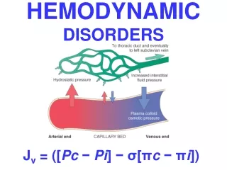
Fluid HomeostasisStarling’s Law Homeostasis is maintained by the opposing effects of: • Vascular Hydrostatic Pressure • and • Plasma Colloid Osmotic Pressure
EDEMA Increased fluid in the interstitial tissue spaces or body cavities. • Increased hydrostatic pressure: – Impaired venous return – Congestive heart failure (poor right ventricular function) – Constrictive pericarditis – Ascites(peritoneal dropsy; e.g. from liver cirrhosis) – Venous obstruction or compression (thrombosis, external pressure, dependency of lower limbs) • Arteriolar dilation (heat; neurohumoral dysregulation) • Reduced plasma osmotic pressure (hypoproteinemia) – Nephrotic syndrome(protein-losing glomerulopathies) – Liver cirrhosis (ascites) – Malnutrition – Protein-losing gastroenteropathy
Lymphatic obstruction – Interstitial fluids are removed via lymphatic drainage, to thoracic duct and left subclavian vein – Inflammation, neoplasm, surgery, irradiation • Sodium retention (water follows sodium) – Excess salt intake with renal insufficiency – Increased tubular reabsorption of sodium (renal hypertension;renal hypoperfusion-- increased renin-angiotensin-aldosterone secretion) • Inflammation (acute, chronic, angiogenesis)
CHF EDEMA • INCREASED VENOUS PRESSURE DUE TO FAILURE • DECREASED RENAL PERFUSION, triggering of RENIN-ANGIOTENSION-ALDOSTERONE complex, resulting ultimately in SODIUM RETENTION
HEPATIC ASCITES • PORTAL HYPERTENSION • HYPOALBUMINEMIA
RENAL EDEMA • SODIUM RETENTION • PROTEIN LOSING GLOMERULOPATHIES (NEPHROTIC SYNDROME)
Transudate vs Exudate • Transudate • results from disturbance of Starling forces • specific gravity < 1.012 • protein content < 3 g/dl, • Exudate • results from damage to the capillary wall • specific gravity > 1.012 • protein content > 3 g/dl,
GENERALIZED EDEMA • HEART • LIVER • KIDNEY Dependent Edemais a prominent feature of Congestive Heart Failure; in legs if standing or sacrum in sleeping patient Periorbital edemais often the initial manifestation of Nephrotic Syndrome, while late cases will lead to generalized edema.
Pulmonary Edema is most frequently seen in Congestive Heart Failure May also be present in renal failure, adult respiratory distress syndrome (ARDS), pulmonary infections and hypersensitivity reactions
Pulmonary Edema • The Lungs are typically 2-3 times normal weight • Cross sectioning causes an outpouring of frothy, sometimes blood-tinged fluid • It may interfere with pulmonary function
Brain Edema • Trauma, Abscess, Neoplasm, Infection (Encephalitis due to say… West Nile Virus), etc The surface of the brain with cerebral edema demonstrates widened gyri with a flattened surface. The sulci are narrowed
Brain Edema Clinical CorrelationThe big problem is: There is no place for the fluid to go! • Herniation into the foramen magnum will kill
SHOCK • Definition: CARDIOVASCULAR COLLAPSE • Common pathophysiologic features: • INADEQUATE CARDIAC OUTPUT and/or • INADEQUATE BLOOD VOLUME • Pathogenesis • Cardiac • Septic • Hypovolemic
GENERAL RESULTS • INADEQUATE TISSUE PERFUSION • CELLULAR HYPOXIA • UN-corrected, a FATAL outcome TYPES of SHOCK • CARDIOGENIC: (Acute, Chronic Heart Failure) • HYPOVOLEMIC: (Hemorrhage or Leakage) • SEPTIC: (“ENDOTOXIC” shock, #1 killer in ICU) • NEUROGENIC: (loss of vascular tone) • ANAPHYLACTIC: (IgE mediated systemic vasodilation and increased vascular permeability)
CARDIOGENIC shock • MI • VENTRICULAR RUPTURE • ARRHYTHMIA • CARDIAC TAMPONADE • PULMONARY EMBOLISM (acute RIGHT heart failure or “cor pulmonale”)
HYPOVOLEMIC shock • HEMORRHAGE, Vasc. compartmentH2O • VOMITING, Vasc. compartmentH2O • DIARRHEA, Vasc. compartmentH2O • BURNS, Vasc. compartmentH2O
SEPTIC shock • OVERWHELMING INFECTION • “ENDOTOXINS”, i.e., LPS (Usually Gm-) • Degraded bacterial cell wall products • Also called “LPS”, because they are Lipo-Poly-Saccharides • Attach to a cell surface antigen known as CD-14 • Gm+ • FUNGAL • “SUPERANTIGENS”, (Superantigens are polyclonal T-lymphocyte activators that induce systemic inflammatory cytokine cascades similar to those occurring downstream in septic shock, “toxic shock” antigents by staph are the prime example.)
Effects of Lipopolysaccharide LPS = lipopolysaccharide TNF = tumor necrosis factor IL = interleukin NO = nitric oxide PAF = platelet-activating factor
SEPTIC shock events(linear sequence) • SYSTEMIC VASODILATION (hypotension) • ↓ MYOCARDIAL CONTRACTILITY • DIFFUSE ENDOTHELIAL ACTIVATION • LEUKOCYTE ADHESION • ALVEOLAR DAMAGE (ARDS) • DIC • VITAL ORGAN FAILURE CNS
CLINICAL STAGES of shock NON-PROGRESSIVE • COMPENSATORY MECHANISMS • CATECHOLAMINES • VITAL ORGANS PERFUSED PROGRESSIVE • HYPOPERFUSION • EARLY “VITAL” ORGAN FAILURE • OLIGURIA • ACIDOSIS IRREVERSIBLE • HEMODYNAMIC CORRECTIONS of no use
Morphologic Features of Shock • Brain: ischemic encephalopathy • lung :DAD (Diffuse Alveolar Damage,) • Heart: subendocardial hemorrhages and necrosis • Kidneys: acute tubular necrosis or diffuse cortical necrosis • Gastrointestinal tract: patchy hemorrhages and necrosis • Liver: fatty change or central hemorrhagic necrosis • DIC • MULTIPLE ORGAN FAILURE
CLINICAL PROGRESSIONof SYMPTOMS(linear sequence) • Hypotension • Tachycardia • Tachypnea • Warm skin Cool skin Cyanosis • Renal insufficiency • Obtundance • Death