Download
1 / 11
110 likes | 468 Vues
HOOK Muscle is only biological cell/tissue that can cause rapid, large-scale movement Role of filamentous proteins understood as great and early breakthrough in cell/molecular biology—lots of protein available, (like Hemoglobin). Sliding Filament Model of Muscle Contraction.

E N D
HOOK • Muscle is only biological cell/tissue that can cause rapid, large-scale movement • Role of filamentous proteins understood as great and early breakthrough in cell/molecular biology—lots of protein available, (like Hemoglobin) Sliding Filament Model of Muscle Contraction Larry M. Frolich, Ph.D. April 15, 2010 I normally cover neurons and muscle together as part of unit on movement—see website
Sliding Filament Model of Muscle Contraction OUTLINE • Motor Unit • Muscle Cell Architecture and Function • Sliding Filamentous Proteins • Muscle Force Properties
Muscle Cells and Neurons • are unique to animals • have “excitable” membranes that transmit action potentials • allow for rapid large-scale movements • Motor Unit is one motor neuron plus the muscle cells that it stimulates (or synapses with)--the minimal construct that allows for movement in our body
Muscle cells • Muscle fibers are cells—visible to naked eye as fibers in meat, chicken, fish • Sarcolemma is muscle cell membrane—”excitable” so has action potentials just like neurons • Because cell is large, T-tubules carry action potential—ionic depolarization—into internal parts of cell • Sarcoplasmic reticulum releases calcium which triggers actin-myosin protein filaments to contract Sequence of events Motor Neuron to Muscle contraction at cellular level (from the Brain Top to Bottom) [link]
Muscle cell or muscle “fiber” is composed of myofibrils which contain sarcomeres or contractile “units” Myo- Sarco- (= muscle)
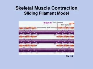
Molecular Basis of Muscle Function • Actin-Myosin “sliding filament” model • Explains • Muscle movement or shortening • Muscle force generation or “contraction” • Actin and myosin filamentous proteins are packed parallel in sarcomeres
How does the actin-myosin complex (sarcomere) shorten and contract the muscle? • Actin = thin filament “lattice-work” • Myosin = thick filament “core” • Ca release triggers the formation of molecular cross-bridges from myosin to actin • Cross-bridges “row” or “reach” for more adjacent binding site on actin.
And the result is muscle movement Put the sliding filaments back into a whole muscle…
Details, details, details… Tropomyosin and troponin create binding site on actin filament Presence of Ca++ exposes binding site “Cocked” cross-bridge on myosin (uses ATP) then attaches to binding site and pulls or “rows” actin filament Cross-bridge linkage is broken and re-cocks to link with next binding site Details Video
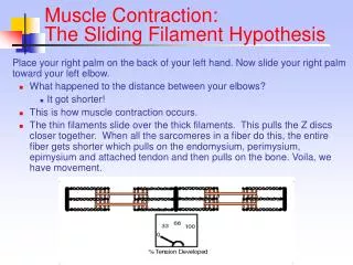
Sliding Filament Model explains Why muscle has peak force at certain length: (ideal actin-myosin overlap for cross-bridge formation)—BUCKET DEMO More muscle cells means more muscle force: (more cross-bridge formation)—EMG, Isolated muscle online lab Concentric/isometric/eccentric contraction: Cross-bridges continue to form and “reach” even if opposing force is greater