Download
1 / 8
140 likes | 3.53k Vues
2. Sliding filament theory of muscle contraction Muscular tissue

E N D
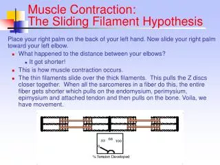
2. Sliding filament theory of muscle contraction • Muscular tissue • Muscular tissue is composed of many elongated cells called muscle fibres (or myoneme) which consist of many myofibril. Each myofibril is composed of two types of proteinaceous myofilaments: actin (thin filament) and myosin (thick filament). • Under the electronic microscope, light and dark bands can be observed in every muscle fibre
The centre of myosin is located at M line while that of actin is located at Z line. Light band is composed of actins while dark band is composed of both actins and myosins. H zone is composed of myosins only.
Myosins are composed of many rod-shaped myoglobins which can be divided into the myosin rod and myosin head. Myosin rods are collected together to comprise the main branch of myosin while myosin heads project out to form the cross-bridges. • Cross-bridges can twist and contain ATPase which hydrolyze ATP to generate energy
Each actin filament (A) is made up of two helical strands of globular actin molecules (G-actin) which twist round each other. It also consists of two accessory proteins called tropomyosin (T) and troponin. Actins and myosin heads can be linked to form the cross-bridges. However, at resting stage, the attachment site is covered by tropomyosin to prevent the formation of cross-bridges. Troponin has high infinity to Ca2+ and can reversibly bind with Ca2+ .
Sarcolemma • The structure of sarcolemma is similar to a typical cell membrane but it is invaginated and surrounds each muscle fibre. Fluid can pass through the membrane and external stimuli can be transmitted through it also.
Sarcoplasm • The cytoplasm of the myofibril is called sarcoplasm and contains a network of internal membranes termed the sarcoplasmic reticulum. Running transverely across the fibre and between fibrils is a system of tubules known as the T system. A T tubule together with a pair of vesicles is called a triad which is involved in the uptake and release of Ca2+ ions and holds 80% of Ca2+ in a muscle cell.
Mechanism of muscle contraction: Nerve impulse is transmitted to sarcolemma Sarcolemma is depolarized Action potential spreads out and reaches the triad through T system Impulse stimulates the release of Ca2+ into the sarcoplasm Ca2+ binds with cross-bridge and changes the conformation of troponin
Attachment site is exposed, ATPase in myosin head is activated to hydrolyze ATP to generate energy Release of energy accompanies cross-bridge formation and pulls the actins towards M line Length of sarcomere is shortened and resulted in muscle contraction Ca2+ is actively pumped back into the triad after excitation, myosin head combines with another ATP Cross-bridge is broken, actins and myosins return to original conformation as in resting stage