Download
1 / 1
20 likes | 179 Vues
STM tip. STM tip. STM tip. STM tip. tunneling. tunneling. tunneling. tunneling. Hole . Hole . Hill. Hill. Metal Phthalocyanines and Porphyrazines Modified Gold Single-Crystal Electrodes Mohammad F. Khanfar 1 , Susan H. Zheng 1 , Hong Zong 2 , Brian M. Hoffman 2 and Sylvie Morin 1

E N D
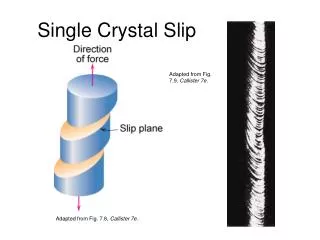
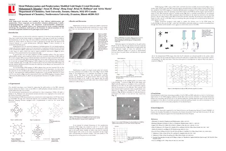
STM tip STM tip STM tip STM tip tunneling tunneling tunneling tunneling Hole Hole Hill Hill Metal Phthalocyanines and Porphyrazines Modified Gold Single-Crystal Electrodes Mohammad F. Khanfar1, Susan H. Zheng1, Hong Zong2, Brian M. Hoffman2 and Sylvie Morin1 1Department of Chemistry, York University, Toronto, Ontario, M3J 1P3 Canada 2Department of Chemistry, Northwestern University, Evanston, Illinois 60208-3113 STM imaging of MPcs such as FePc, CoPc, and CuPc have been carefully characterized by Hipps and co-workers [5]. In their work some MPc molecules appear with dark (holes) or bright regions in their centers. They have proposed that STM imaging of adsorbed phthalocyanines monolayers occurs through orbital mediated tunneling mechanism, where the tunneling current flows between the STM tip and the organic layer beneath via empty or half filled molecular orbital of suitable energy. Fig. 12 is a schematic presentation of the proposed mechanism [9,10]. In the three MPcs mentioned above, the orbital of the metal ion involved in the tunneling process is the dz2. That orbital is half filled in both CoPc and FePc, therefore, the metal ionic center appears as a bright. In CuPc, the dz2 is full filled, hence no tunneling takes place through such an orbital and the metal ionic center appears as a hole. Similar theoretical treatment could apply to explain the features seen in the STM image for C24H24N8S8Mg. In this case the Mg2+ center has a full filled pz molecular orbital, as a result no tunneling flows through that orbital and the metallic center appears as a hole in the center of the MPz. Abstract Gold single-crystal electrodes were modified by three different phthalocyanines and porphyrazines. These compounds are hexadecafluro ruthenium phthalocyanine, F16RuPc, octapropylporphyrazine, C40H58N8, and magnesium octakis(methylthio) porphyrazine, C24H24N8S8Mg. The modified surfaces were characterized by in-situ scanning tunneling microscopy. Our preliminary results point to the formation of ordered monolayers of the mentioned compounds adsorbed on the gold single-crystal surfaces. 3.Results and Discussion Modification of the Au (111) surface by F16RuPc is presented in Fig 6. The adsorbed phthalocyanine molecules are seen as bright spots that are decorating all six Au (111) terraces visible in the figure. Figure 12: Electron tunneling through porphyrazines modified gold single crystal surfaces (adapted from Ref. 10). 1.Introduction Phthalocyanines are macrocyclic molecules composed of four fused iminoisoindoline units. They have central cavities large enough to accommodate a wide range of metal ions. In addition, functionalization of the four rings with many different substituents has been reported in a large number of articles. Nature of metal ion as well as substituents strongly affects the physical and chemical behavior of a metal phthalocyanine (MPc)[1]. Figure 1 shows structure of an unsubstituted phthalocyanine. Porphyrazines, Pz’s, are structural analogues of phthalocyanines. Pz’s are simply porphyrin compounds with four nitrogen atoms at the meso positions. A significant attention has been devoted to Pzs and Metal Pzs (MPzs), mainly due to convenience of preparation of these macrocycles with a wide range of metal central ions and/or organic peripheral substituents. Figure 2 shows structure of the unsubstituted free base porphyrazine [2]. In general there are two main categories of MPcs modified surfaces. The first involves polycrystalline surfaces, such as gold and carbon, which are covered by thin films of MPcs [3]. In the second category, MPcs exist as ordered monolayers adsorbed on single-crystal electrodes, such as Au (111) and Cu(100) [4]. MPcs adsorbed molecules can be easily recognized when monolayers of 25–250 nm2 are imaged. STM imaging of MPcs adlayers has been performed under ultra high vacuum conditions [5], in air [6], and in solutions [7]. It has been found that STM images can provide valuable information about orientation the molecules in the adlayers and dimensions of the adsorbed molecules [8]. To our knowledge, STM imaging of MPzs adlayers have not been reported. We are also interested in the investigation of the electrochemical behavior of Pzs monolayers. As a first step in this direction we have modified gold single-crystal surfaces with two Pzs, octapropylporphyrazine, C40H58N8, and magnesium octakis(methylthio) porphyrazine, C24H24N8S8Mg. Structures of these two Pz’s are shown in Fig. 3 and 4, respectively. In this work STM imaging of hexadeca fluoro ruthenium (II) phthalocyanine, F16RuPc was also performed. Structure of F16RuPc is shown in Fig. 5. Figure 6: (a) STM image of F16RuPc adlayer on Au (111) at 0.15 VSCE. The image was acquired in 0.1 M HClO4. Set point current and tip bias of the W tip were 1 nA and -0.050 V, respectively. (b) constant current contour along the white line indicated in part (a). Figure 8: STM image of C40H58N8 adlayer on Au (111) at 0.15 VSCE. The image was acquired in 0.1 M HClO4. Set point current and tip bias of the W tip were 1 nA and -0.150 V, respectively. Molecules appear to be laying flat on the gold surface. Height of the Pz molecules and distances between molecules were estimated from Fig. 8. Fig. 9 shows a height profile that allows molecular height of three Pz molecules to be estimated. Molecular height of C40H58N8 is approximately 0.09 nm above the gold surface and molecules are separated by ca. 1 nm. All of the demonstrated STM images show that the Pz molecules are oriented parallel to the surface rather than vertically. The intermolecular distance between Pz molecules is smaller than what is expected for a end-to-end packing of the alkyl side chains. That observation points to interdigitation of adjacent alkyl side chains, as proposed in Fig. 13. Modification of gold (111) single crystal surface by C40H58N8 has been performed successfully in this work. Fig. 7 shows STM image of the porphyrazine as a monolayer decorating the single-crystal surface. The figure shows how porhyrazines molecular domains are distributed on the gold terraces. Drift in the image prevents all domains to be visible. It also appears that the gold reconstruction is lifted (at least partially) under these conditions (gold islands are visible in Fig. 7). Figure 9: Molecular height of C40H58N8 molecules above the gold surface STM image of the modified gold (111) surface with C24H24N8S8Mg is shown in Fig. 10. In this image, each MgPz molecule appears as a doughnut with a central hole. Similarly to the other Pz the gold reconstruction appears lifted by the presence of the molecules. This contrast with layers formed with Pc molecules where no gold islands were observed (see Fig. 6 for comparison). A cross section analysis of Fig. 10 allows estimation of molecular distances and height to be about 1.5 nm and 0.05 nm, respectively (Fig. 11). 2. Experimental The adsorbed monolayers were formed by immersing the gold surfaces in the MPc saturated benzene solution for almost 1 hour, or the MPzs 0.1 mM methylene chloride solutions were used and the immersion time was 24 hours. The electrochemical measurements were performed in a three compartment Teflon cell under a blanket of N2. Platinum wire and saturated calomel electrodes were employed as the counter and the reference electrodes, respectively. The electrochemical STM measurements were carried out at room temperature, with W tips etched in 2 M NaOH. The tips were coated with Apiezon wax to minimize residual faradaic currents. A Molecular Imaging Picoscan equipped with a bi-potentiostat is used for the in-situ STM experiments. Figure 13: Interdigitation of adjacent MPz molecules on gold (111) surface 4.Conclusions Our preliminary results show interesting assembly of the Pz, MPz and MPc molecules on Au(111). In the future, improvement of the STM imaging conditions should allow more accurate evaluation of molecular dimensions, domain size/orientation and 2D unit cell of the adsorbed molecules. Another important aspect of this work is the investigation of the effects of electrode potential on molecular packing, domain size and on orientation of the adsorbed layers as well as on the redox properties of the adsorbed layers. Acknowledgments This work was financially supported by the Natural Sciences and Engineering Research Council (NSERC) of Canada, the Canadian Foundation for Innovation, the Ontario Innovation Trust and York University. S. M. also acknowledges the financial support from the Canada Research Chair Program. Figure 7: STM image of C40H58N8 adlayer on Au (111) at 0.15 VSCE. The image was acquired in 0.1 M HClO4. Set point current and tip bias of the W tip were 1 nA and -0.150 V, respectively. • References • 1 Bekaroglu, O., Journal of Porphyrins and Phthalocyanines 2000, 4, 465-473. • 2 Rodriguez-Morgade, M. Salome; S., Pavel A. J. Porphyrins. Phthalocyanines 2004, 8, 1129-1165. • 3 Lippel, P. H., Wilson, R. J., Miller, M. D., Woll, C., Chiang, S., Phys. Rev. Lett. 1989, 62, 171-174. • 4 Miao, P., Robinson, A. W., Palmer, R. E., Kariuki, B. M., and Harris, K. D. M., J. Phys. Chem. B 2000, 104, 1285 – 1291. • 5 Barlow, D; Scudiero, L; and Hipps, K, W; Ultramicroscopy 2003, 97, 47-53. • 6 Lie, S. B.; Yin, S. X;Wang, N. H; Xi, F; Lui, H. W; Xu, B; Wan, L. J; and Bai, C. L; J. Phys. Chem. B 2001, 105, 10838-10841. • Yashimoto, S; Tada, A; Suto, K; Yau, S-L.; and Itaya, K., Langmuir 2004, 20, 3159 – 3165. • 8 Yashimoto, S; Tada, A; Suto, K; and Itaya, K; J. Phys. Chem. B 2003, 107, 5836-5843. • 9 Scanning Tunneling Spectroscopy, by K W Hipps; a chapter in "Handbook of Applied Solid State Spectroscopy", Ed: Vij, D. R., Kluer Scientific (2005). • 10 Lu, X.; and Hipps, K. W. J. Phys. Chem. B 1997, 101, 5391-5396. Figure 1: phthalocyanine Figure 2: porphyrazine Figure 3: octapropylporphyrazine, C40H58N8 Figure 10: 40 x 40 nm2 STM image of C24H24N8S8Mg in 0.1 M HClO4. Tip bias and set point are -0.135 V and 1.450 nA, respectively. In an attempt to estimate dimensions of the porphyrazine molecules, STM image was zoomed in and 10 x 10 nm2 image analyzed (see Fig. 8). Each Pz molecule can be recognized as a fuzzy spot on the gold surface. Ideally, we believe that each Pz molecule should appear with a hole at its center surrounded by four bright spots corresponding the aromatic residues. The poor quality of the image prevent us at this point to see such details. Figure 11: Molecular height of C24H24N8S8Mg molecules above the gold surface. Figure 4: Magnesium octakis(methylthio)- Porphyrazine, C24H24S8N8Mg Figure 5: hexahalo ruthenium(II) phthalocyanine