Download
1 / 13
160 likes | 271 Vues
Explore the histology of pituitary, thyroid, and parathyroid glands, including detailed structures and cell types found within each gland. Discover the function and composition of adenohypophysis, neurohypophysis, thyroid follicles, and more.

E N D
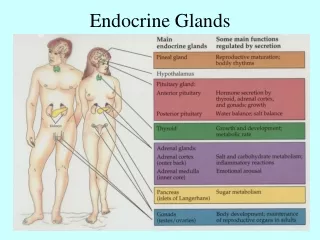
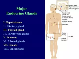
Pituitary gland • Pituitary gland- magnified • Thyroid and parathyroid glands • Thyroid follicles- magnified
Pituitary gland Intraglandular cleft Neurohypophysis Adenohypophysis Intermediate part
Neurohy-pophysis Intermed-iate part Pituitary gland- magnified Adenohypophysis Intragla-ndular cleft Chromo-phobes Chrom-ophils A vesicle Sinusoid Pitucyte
Thyroid and parathyroid glands Colloid Chief cells Parafollicular cells Follicular cells Sinusoid Parathyroid Thyroid follicles
Thyroid follicles- magnified Colloid Follicular cells Parafolli-cular cells
Pituitary gland: • It has 2 major parts: adenohypophysis and neuropypophysis. • Adenohypophysis: • There are 3 parts in adenohypophysis: Pars distalis, pars intermedia and pars tuberalis. • Pars distalis: • It is the largest subdivision of the gland. It is composed of glandular cells arranged in irregular cords or clumps close to the blood capillaries and sinusoids. It has: • Chromophobes: • These make 50% of cell population. These are small cells with lightly stained cytoplasm and pale nucleus. These cells are found in groups.
2. Chromophils: Larger cells with granular cytoplasm. They are of two types based on their staining properties. a. Acidophils (alpha cells): They contain eosinophilic granules and secrete growth hormone and lactogenic hormone b.Basophils (beta cells): Secretory granules in the cytoplasm are basophilic in nature. These cells secrete thyroid stimulating hormone, follicle stimulating hormone, luteinizing hormone and adrenocorticotrophic hormone.
Pars intermedia: This part is seen between the neurohypophysis and pars distalis. The cells here are arranged in the form of vesicles. These are low columnar basophilic cells with colloid in their cytoplasm. They secrete melanocyte-stimulating hormone. Between the pars intermedia and pars distalis there is a cavity called itnraglandular cleft. Pars nervosa: It consists of terminal parts of axons of secretory neurons whose cell bodies are in the hypothalamus. In between the nerve fibers there are neuroglial cells called pituicytes, which are variable in size. Herring bodies, which are local accumulations of the neurosecretory substances in the axoplasm of the nerve fibers, are also seen here.
Thyroid gland: A capsule consisting of fibroelastic connective tissue sends septa into the gland, providing internal support and carrying blood vessels, nerves and lymphatics into its substance. These septa divide the gland into masses of irregular form and are often called lobules. The thyroid gland is consisting of two types of secretory cells, the cells of the thyroid follicles (follicular cells) and parafollicular cells. Thyroid follicles: Each thyroid follicle is a closed, hollow, spherical structure containing a yellow, viscid, semi fluid material called colloid, which stains pink with eosin.
A single layer of cuboidal epithelium that is supported by a basement membrane lines the follicles. The follicles are separated from each other by a highly vascular connective tissue. Para follicular cells (light cells or C cells) These are large polyhedral or oval cells situated singly or in small groups between the follicles of the thyroid gland. They are always separated from the lumen of the follicles by the follicular cells. However, like the follicular cells they are situated on the inner aspect of the basement membrane that surrounds the follicle. In ordinary sections they have pale cytoplasm.
The parathyroid gland: A thin connective tissue capsule covers the gland. Septa along with blood vessels and nerve fibers pass into the gland from the capsule. There are mainly two types of cells in the gland. Principal cells: These are the main type of cells seen in the gland. These cells are of three categories Dark Principal cells: They have dark staining cytoplasm and are most active cells
Light Principal cells: These are cells with pale cytoplasm Clear Principal cells: These cells are not easily stained Oxyphils: These are larger cells found in clumps. They have abundant cytoplasm, which takes eosin stain and a small nuclei, which take darker stain.