Download
1 / 12
120 likes | 220 Vues
Explore the latest advancements in Hadron Therapy detectors and their specifications for precise and effective treatment delivery. Understand the importance of physical data in dose distribution and modeling for optimal patient care.

E N D
Hadron Therapy RequirementsDetectors: emerging rationale & specificationsChris Moore Chris Moore, CMPE, 19 March 2014
Chris Moore, CMPE, 19 March 2014 RADIOTHERAPY STATUS CHECK Today’s ‘dose hard, fast & tight’ paradigm Importance of physical data. Planning 3D & 4D pre-treat CT reduction. Body, organs & disease derived. Dose distribution & effect modelled. In-Treatment Dynamic dose-shaping. kV & MV Image guidance. In-vivo dosimetry. Measure Meaurement: Certainty Adapt inter-#
Adapt Control? Change Scanning beam Adapt Control? Chris Moore, CMPE, 19 March 2014 PROTON SCANNING BEAM THERAPY Planned (simulated) vs Actual (measured) Lomax, Oxford, 2008.
Chris Moore, CMPE, 19 March 2014 Machine Commissioning Daily Machine QA PBS Dose QA
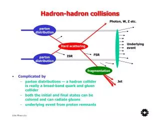
INTO-ROOM: Model April 2013Varian & PSI collaboration motion management for p-therapy of lung,liver, pancreas & breast, using a 4-D PTP simulator developed by PSI. von Siebenthal et al ETH & PSI 4D-CT from 4D-MRI IN-ROOM: Indirect Passive Activ’n PET*** P-Radiograph* MV CT** IN-ROOM: Direct Active Synchronous Ranging/Adaptation Moore, Price & Parkhurst, Christie, Manchester Chris Moore, CMPE, 19 March 2014 UNCERTAINTIES MOTION, SHRINKAGE etc. "The machine is not the problem“ "The problem is the patient." Engelsman mms to cms WHERE BEAM, DOSE WENT "The advantage of protons is that they stop. The disadvantage…we don't know where..." Range Uncertainties: Set-up errors CT number-to-proton stopping power, Inter-fraction anatomy change intra-fraction motion Engelsman mms to cms, 5-20% CONTROL STRATEGIES Re-scanning averaging/smear Re-tracking to repeatedly follow tumour Prompt-gamma ranging P-tomography Lomax & de Water mms, 5% *Uwe Schneider, Zurich Alexander Tourovsky, PSI ***F Attanasi et alPhys. Med. Biol. 56 5079 **Ospedale San Rafaele, Milan Francesca Albertini, PSI
Chris Moore, CMPE, 19 March 2014 PTCOG-52, June 2013, Essen, Engelsman; 1. TODAY; roll-out of cone-beam (CB) or diagnostic CT. 2. 5 YRS; have clinical experience in routine, full 3D/4D imaging, along with acquisition & matching. - Online evaluation of plan accuracy - ‘Plan of the day’ using in-room (CB)CT *CMPE MRC grant 3. 10 YRS; automated on-line re-planning in 10 secs, i.e. while patient is on treatment couch. - Automation at a level effectively removing experts from treatment prep process. - Needs (too?) much faith in RT dose planning. - Consequently, in vivo dosimetry is a necessity Replacing off-line dosimetric plan-specific QA.
Chris Moore, CMPE, 19 March 2014 (Moore, Price & Parkhurst, Christie, Manchester) Entrance surface moving & deforming. Tissues at depth moving & deforming. Tumour at depth moving, deforming. Limited correlation for prediction.
Chris Moore, CMPE, 19 March 2014 DRIVING FORCES: verify & alert ‘‘There is a lack of consensus about every issue in radiotherapy including what are significant complications’’ Fletcher GH. Hypofractionation: Lessons from Complications’, Radiother Oncol 20, 105, 1991 Photons Friberg & Ruden, Hypofractionation in radiotherapy. An investigation of injured Swedish women, treated for cancer of the breast, Acta Oncol, 48, 822-31, 2009 ’disregarded telangiectasia, ulceration of the skin, hyperpigmentation.’ ‘focused our interest on brachial plexus injuries’: Protons Dr Brooks, Director prostate & colorectal cancers, American Cancer Society. No evidence protons do a better job for prostate & colorectal cancer. Some studies suggest side effects might be higher. 1. Urinary (bladder problems) 2. gastrointestinal (rectal leakage and bleeding) 3. Sexual (erectile dysfunction).
Adapt Control? Change Scanning beam Adapt Control? Chris Moore, CMPE, 19 March 2014 DOSIMETER USE Beam/spot Phantom surface Phantom volume Body surface/entrance Tumour implant e.g. lung, breast Body cavities e.g. mouth, sinus OARs ‘sentinel’ implant e.g. spine, skull base, eye
Chris Moore, CMPE, 19 March 2014 • DETECTORS & SPECS FOR RADIATION SOURCES • Bremstrahlung, orthovoltage X-ray tube photons, typically 200-300 kVp • MeV X-ray photons from linear accelerators, typically 4-20 MeV • ~ 60 MV proton cyclotron ‘superfiicial’ source c.f. at Clatterbridge • ~250 MV proton source at National Proton Treatment Centre (2018). • ~350 MV proton imaging source; emerging. • Sensitivity • Energy dependence • Linearity • Saturation • Readout time • Consistency/reproducibility • Long term stability. • Dose Integration & Dose Rate • Planar & volume micro-arrays • Tissue equivalence • Encapsulation effects • Configurable Detector Array (I suggest)
Chris Moore, CMPE, 19 March 2014 • DETECTORS & SPECS FOR RADIATION SOURCES • ~ 60 MV proton cyclotron ‘superfiicial’ source c.f. at Clatterbridge • ~250 MV proton source at National Proton Treatment Centre (2018). • ~350 MV proton imaging source; emerging. • QA, proton beam spot ~3mm sigma suggests 2D/3D arrays - cGy to Gy capability • (at isocentre can be 6-9mm) - ~0.1 mm resolution • - >1 cm areal array • - >1 cc volume array • - <0.01 sec • Body entrance, central, gradient & far depth field use - cGy to Gy capability • (energy change <0.1 sec @ PSI-G2) - ~ 0.1 mm resolution • - > 100 sq.cms • - > 1000 cu.cms • - ~ 1mm3 implant • - <0.01 sec • NB. Micro-mesh Micromegas, UoPenn, resolution 1.1 mm (1σ), 1 ms time resolution. • GEM devices offer 0.1mm pixels.
Chris Moore, CMPE, 19 March 2014 Close Medical aspiration since the 1960s!