Download
1 / 1
20 likes | 186 Vues
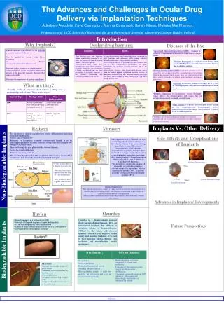
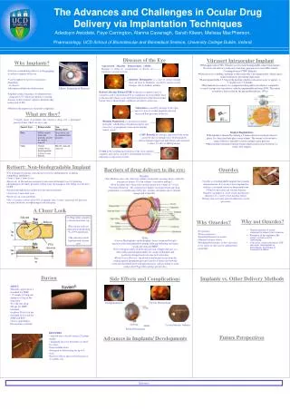
The Advances and Challenges in Ocular Drug Delivery via Implantation Techniques. Silicone cup containing drug. EFFECT OF ENVIRONMENT AND CLOZAPINE ON BASAL AND STIMULATED MEDIAL PREFRONTAL GABA RELEASE IN TWO RAT MODELS OF SCHIZOPHRENIA. Top View. Release orifice. 3mm.

E N D
The Advances and Challenges in Ocular Drug Delivery via Implantation Techniques Silicone cup containing drug EFFECT OF ENVIRONMENT AND CLOZAPINE ON BASAL AND STIMULATED MEDIAL PREFRONTAL GABA RELEASE IN TWO RAT MODELS OF SCHIZOPHRENIA Top View Release orifice 3mm AdedoyinAwodele, Faye Carrington, AlannaCavanagh, Sarah Kileen, Melissa MacPherson. Pharmacology, UCD School of Biomolecular and Biomedical Science, University College Dublin, Ireland. Side View PVA structure tab 2mm 5mm Diseases of the Eye • Vitrasert Intraocular Implant • FDA approved in 1996, Vitrasert was first non-biodegradable, intravitreal implant. • Used for the delivery of the anti-viral drug, ganciclovir to treat AIDs-related Cytomegalovirus (CMV) Retinitis. • Ganciclovir is a synthetic analogue of the nucleoside 2-deoxyguanosine, which causes chain termination, preventing replication. • Each implant holds 4.5-5mg of the prodrug which is released at a rate of approx. 1-1.5µg/hr over 5-8 months. • The 4mm device consists of a compressed drug pellet core which is completely covered, except at its top surface, with the impermeable polymer; EVA. This entire assembly is then coated by the permeable polymer; (PVA). Why Implants? • Age-related Macular Degeneration (AMD) : Damage to retina by accumulation of drusen can cause loss of central vision. • Provide sustained drug delivery to the posterioror anterior segment of the eye • Can be applied to various ocular layers depending • on disease: subconjuctival/intravitreal/intrasceral • Implants reduce frequency of administration • compared to I.V. Route in relation to treating diseases of the posterior segment, therefore they reduce risk of SEs • Minimise the importance of patient compliance. • Diabetic Retinopathy is a type of retinal damage that can lead to blindness caused by microvascular changes due to diabetes mellitus. Above: Insertion of Retisert Diabetic Macular Edema (DME) is the most common cause of visual loss and is characterised by accumulation of extracellular fluid in the macular which occurs after the break down of the blood-retinal barrier due to dilated hyper-capillaries and micro-aneurysms.. • Glaucoma is caused by damage to the optic nerve by loss of retinal ganglion cells and increased fluid pressure in the eye. What are they? • .Usually made of polymers that release a drug over a prolonged period of time. There are two types: • Retinitis Pigmentosais a progressive retinal dystrophy which affects the photoreceptors and causes loss of peripheral vision and eventually central vision. • Surgical Implantation: • The implant is inserted by making a 5-6mm scleral incision into the pars plana. It is then fixed into place using sutures. The wound is closed and a saline solution is injected to restore normal ocular pressure. • Most patients experience blurred vision which usually clears between 2-4 weeks after surgery. • CMV Retinitis is a chronic, infection of the retina caused by the cytomegalovirus. Predominantly affects immunosuppressed individuals and estimated to affect 15-40% of AIDS patients. Uveitis is the swelling and irritation of the uvea (anterior segment) and can be caused by autoimmune disorders, infection or exposure to toxins. • Retisert:Non-biodegradable Implant Barriers of drug delivery to the eye: Ozurdex • For treatment of chronic, non-infectious uveitis (inflammation) including sympathetic ophthalmia. • 3mm x 2mm x 5mm in size • Reservoir of flucinoloneacetonide (corticosteroid thought to act by inducing phospholipase A2 inhib. proteins). 600ng a day decreasing to 300-400ng over the first month. • Inserted through the pars plana into the vitreous humour • Active for 2 and a half years • Removal can cause problems • SEs = Cataracts (observed in 90% of patients after 3 years), increased I.O pressure, eye pain, headache, nasopharyngitis and joint pain. Dynamic: • Tear Dilution- max. only 30µl tear volume comfortably accomm. In eye when the average eye drop is 50 µl in volume – inevitable spillage! • Naso-lacrimal duct- when tears exceed normal tear volume of 7-10 µl • Systemic Removal – the conjunctiva is highly vascularised and any drug permeating it is rapidly removed by the systemic circulation and eventually transported to the GIT. Ozurdex is a biodegradable implant that contains demaxethasone. It is an intravitreal implant that delivers a sustained release of demaxaethasone (700ml) to the retina and vitreous humour. Ozurdex can improve visual acuity and macular thickness. It is used to treat macular edema, Retinal vein occlusion and non-infectious uveitis (posterior). A Closer Look • 0.59mg tablet is held in a silicone elastomer cup. • The release orifice is separated from the drug • by a PVA membrane. • The structure is held together with silicone glue • Why not Ozurdex? • Why Ozurdex? • Shorter duration of action compared to retinal vein occlusion. • Retention of the implant in the correct position can be challenging. • Can cause cataract formation, IOP elevation, subconjuntivalhemorrhage, hyperemia or conjunctivaledema. • It’s potency • Dose consistency • Extended Duration of action • Minimal adverse effects • Biodegradable nature- It does not need to be retrieved and can be administered repeatedly. • Static: • Cornea-Hydrophobic and hydrophilic layers connected by tight junctions and containing efflux pumps inducing multidrug resistance. (p-glycoprotein and MRP) • Sclera-Opaque matrix of proteoglycans and collagen that acts as a filter with preferred permeability for small, hydrophilic and positively charged molecules due to it’s structure. • Blood-Ocular Barriers –the blood-retinal barrier arises from the retinal pigment epithelium prevents transfer of molecules between itself and choroidal blood with tight junctions and according to some studies show P-gp efflux pumps present also. Iluvien Implants vs. Other Delivery Methods Side Effects and Complications • ABOUT • Recently approved as a treatment for DME • It weighs 0.18mg and dispenses 0.2µg of the drug daily • It is the only drug therapy for DME treatment • In phase II trials for the treatment of wet and dry AMD and RVO • Active ingredient is fluocinoloneacetonide Vitreous Haemorrhage Endophthalimitis Cystoid Macular Oedema Retinal Detachment • DELIVERY • injected intra-vitreally using a 25 gauge needle. • minimally invasive procedure, no need for suture • Non-erodible insert • Designed to deliver drug for up to 3 years • Easier to deliver then retisert because of its smaller size Future Perspectives Advances in Implants/ Developments References