Download
1 / 33
340 likes | 587 Vues
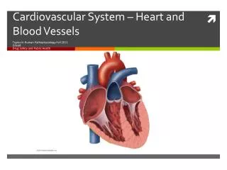
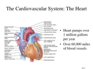
Chapter 20 THE CARDIOVASCULAR SYSTEM: THE HEART. Lecture Outline. INTRODUCTION. The cardiovascular system consists of the blood, heart, and blood vessels. The heart is the pump that circulates the blood through an estimated 60,000 miles of blood vessels.

E N D
Chapter 20THE CARDIOVASCULAR SYSTEM: THE HEART Lecture Outline
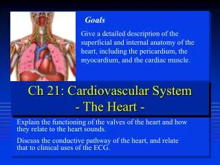
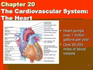
INTRODUCTION • The cardiovascular system consists of the blood, heart, and blood vessels. • The heart is the pump that circulates the blood through an estimated 60,000 miles of blood vessels. • The study of the heart and diseases associated with it is known as cardiology. Principles of Human Anatomy and Physiology, 11e
Location of the heart • The heart is situated between the lungs with about two-thirds of its mass to the left of the midline. • It is about the size of your closed fist. Principles of Human Anatomy and Physiology, 11e
Pericardium • The heart is enclosed and held in place by the pericardium, a membrane. • It confines the heart in its position in the chest • It also allows movement during contraction • An inflammation of the pericardium is known as pericarditis. It is caused by a virus and causes pain. Treatment includes draining fluid through a needle. Principles of Human Anatomy and Physiology, 11e
Layers of Heart Wall • The wall of the heart has 3 layers: • Epicardium • Thin, transparent outer layer • Myocardium • cardiac muscle layer is the bulk of the heart, it does the pumping • Endocardium • Very thin, lines the chambers and vessels Principles of Human Anatomy and Physiology, 11e
Chambers and Sulci of the Heart • Four chambers • 2 upper atria (right and left atrium) • 2 lower ventricles (right and left) • Sulci - grooves on surface of heart containing coronary blood vessels and fat Principles of Human Anatomy and Physiology, 11e
Right Atrium • Blood from the body returns to the heart and goes into the right atrium. • This is shown as blue because the oxygen has been used up • Receives blood from 3 sources • superior vena cava, inferior vena cava and coronary sinus Principles of Human Anatomy and Physiology, 11e
Right Ventricle • Blood from the right atrium then flows into the right ventricle • Blood leaves the right ventricle through pulmonary valves, it then goes into the pulmonary artery to pick up oxygen Principles of Human Anatomy and Physiology, 11e
Left Atrium • Forms most of the base of the heart • Shown as red because it has oxygen • Receives blood from lungs through 4 pulmonary veins Principles of Human Anatomy and Physiology, 11e
Left Ventricle • Forms the apex (bottom) of heart • It has oxygenated blood (red) • Blood from the left atrium flows through the left ventricle and into the aorta, where it is pumped through the body. Principles of Human Anatomy and Physiology, 11e
Myocardial Thickness and Function • Thickness of myocardium varies according to the function of the chamber • Atria are thin walled, deliver blood to adjacent ventricles • Ventricle walls are much thicker and stronger • right ventricle supplies blood to the lungs (little flow resistance) • left ventricle wall is the thickest to supply systemic circulation
HEART VALVES AND CIRCULATION OF BLOOD • Valves open and close in response to pressure changes as the heart contracts and relaxes. • Atrioventricular (A/V) • Semilunar Principles of Human Anatomy and Physiology, 11e
Atrioventricular Valves Open • A-V valves open and allow blood to flow from atria into ventricles when ventricular pressure is lower than atrial pressure Principles of Human Anatomy and Physiology, 11e
Atrioventricular Valves Close • A-V valves close preventing backflow of blood into atria Principles of Human Anatomy and Physiology, 11e
Semilunar Valves • SL valves open with ventricular contraction • allow blood to flow into pulmonary trunk and aorta • SL valves close with ventricular relaxation • prevents blood from returning to ventricles, blood fills valve cusps, tightly closing the SL valves Principles of Human Anatomy and Physiology, 11e
Flow of Blood • Oxygen enters the body through the lungs • Pulmonary veins carry it to the left atrium (red) • It then goes through the left ventricle (red) • The aorta pumps it into the body through arteries (red) • The body uses up the oxygen (red to blue) • Blood returns to the heart through veins (blue) • It enters the right atrium (blue) • It goes through the right ventricle (blue) • Then it goes into pulmonary arteries to get more oxygen from the lungs and the process starts over (blue to red) Principles of Human Anatomy and Physiology, 11e
Blood Circulation • Blood flow • blue = deoxygenated • red = oxygenated • Heart arteries arterioles capillaries venules veins heart Principles of Human Anatomy and Physiology, 11e
Project Part 1 • Get a poster. Divide it in half. • On today’s half, draw and color a picture of the heart. • Refer to page 687. • Include the following: • Both atria and ventricles • Vena cava • Aorta • Pulmonary arteries • Pulmonary veins Principles of Human Anatomy and Physiology, 11e
Autorhythmic Cells: The Conduction System • Cardiac muscle cells are autorhythmic cells because they are self-excitable. They repeatedly generate spontaneous action potentials that then trigger heart contractions. • These cells act as a pacemaker to set the rhythm for the entire heart. • They form the conduction system, the route for propagating action potential through the heart muscle. Principles of Human Anatomy and Physiology, 11e
Electrocardiogram • A recording of the electrical changes that accompany each cardiac cycle (heartbeat) is called an electrocardiogram (ECG or EKG). • The ECG helps to determine if the conduction pathway is abnormal, if the heart is enlarged, and if certain regions are damaged.
THE CARDIAC CYCLE • A cardiac cycle consists of the systole (contraction) and diastole (relaxation) of both atria, rapidly followed by the systole and diastole of both ventricles. • Pressure and volume changes during the cardiac cycle • This is what is measured when blood pressure is taken. Principles of Human Anatomy and Physiology, 11e
Auscultation • The act of listening to sounds within the body is called auscultation, and it is usually done with a stethoscope. • The first heart sound (lubb) is created by blood turbulence associated with the closing of the AV valves. • The second heart sound (dupp) represents the closing of the semilunar valves. Principles of Human Anatomy and Physiology, 11e
CARDIAC OUTPUT • Cardiac output (CO) is the volume of blood ejected from the left ventricle into the aorta each minute. • Cardiac reserve is the ratio between the maximum cardiac output a person can achieve and the cardiac output at rest. Principles of Human Anatomy and Physiology, 11e
Regulation of Heart Rate • Nervous control from the cardiovascular center in the medulla • Heart rate is also affected by hormones • And by ions (Na+, K+, Ca2+) • And by age, gender, diet, physical fitness, and temperature Principles of Human Anatomy and Physiology, 11e
Clinical Problems • MI = myocardial infarction • death of area of heart muscle from lack of O2 • replaced with scar tissue • results depend on size & location of damage • Blood clot • use clot dissolving drugs streptokinase or t-PA & heparin • balloon angioplasty Principles of Human Anatomy and Physiology, 11e
Risk Factors for Heart Disease • Risk factors in heart disease: • high blood cholesterol level • high blood pressure • cigarette smoking • obesity & lack of regular exercise. • Other factors include: • diabetes • genetic predisposition • male gender • high blood levels of fibrinogen (blood clotting factor) • left ventricular hypertrophy Principles of Human Anatomy and Physiology, 11e
Desirable Levels of Blood Cholesterol for Adults • TC (total cholesterol) under 200 mg/dl • LDL under 130 mg/dl • HDL over 40 mg/dl • Normally, triglycerides are in the range of 10-190 mg/dl. • Among the therapies used to reduce blood cholesterol level are exercise, diet, and drugs. Principles of Human Anatomy and Physiology, 11e
EXERCISE AND THE HEART • A person’s cardiovascular fitness can be improved with regular exercise. • Aerobic exercise (any activity that works large body muscles for at least 30 minutes, preferably 3 – 5 times per week) increases cardiac output and elevates metabolic rate. • Regular exercise also decreases anxiety and depression, controls weight. Principles of Human Anatomy and Physiology, 11e
Coronary Artery Disease • Heart muscle receiving insufficient blood supply • Caused by narrowing of vessels • Risk factors for development of CAD include high blood cholesterol levels, high blood pressure, cigarette smoking, obesity, diabetes, “type A” personality, and sedentary lifestyle. • Treatment • drugs, bypass graft, angioplasty, stent Principles of Human Anatomy and Physiology, 11e
Arrhythmia • Arrhythmia (disrhythmia) is an irregularity in heart rhythm resulting from a defect in the conduction system of the heart. Principles of Human Anatomy and Physiology, 11e
Congestive Heart Failure • Congestive heart failure is a chronic or acute state that results when the heart is not capable of supplying the oxygen demands of the body. • Causes of CHF • coronary artery disease, hypertension, valve disorders, congenital defects • Left side heart failure • less effective pump so more blood remains in ventricle • heart is overstretched & even more blood remains • blood backs up into lungs as pulmonary edema • suffocation & lack of oxygen to the tissues • Right side failure • fluid builds up in tissues as peripheral edema Principles of Human Anatomy and Physiology, 11e
Murmurs • A heart murmur is an abnormal sound that consists of a flow noise that is heard before, between, or after the lubb-dupp or that may mask the normal sounds entirely. • Some murmurs are caused by turbulent blood flow around valves due to abnormal anatomy or increased volume of flow. • Not all murmurs are abnormal or symptomatic, but most indicate a valve disorder. Principles of Human Anatomy and Physiology, 11e
Project Part 2 • On the other side of your poster, you will describe a cardio vascular disease or disorder. • Include the following: • Name of disease • Type of disease (genetic, dietary etc) • Risk Factors • Population Affected • Signs/Symptoms • Treatments • Prognosis • Disease by Last Name: • A = Heart Murmurs • B → = Arrhythmias • C D = Heart Attack (MI) • F K = Hypertension • L M = Coronary Artery Disease • N P = Arteriosclerosis • R S = Congenital Heart Defects Principles of Human Anatomy and Physiology, 11e