Download

1 / 66
660 likes | 836 Vues
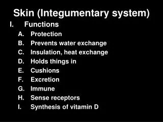
Skin and the Integumentary System. Chapter 6. What exactly is skin?. It is an organ composed of several kinds of tissues Largest organ Performs many functions Maintains Homeostasis Prevents harmful substances such as chemicals and microorganisms from entering body Prevents water loss

E N D
Skin and the Integumentary System Chapter 6
What exactly is skin? • It is an organ composed of several kinds of tissues • Largest organ • Performs many functions • Maintains Homeostasis • Prevents harmful substances such as chemicals and microorganisms from entering body • Prevents water loss • Maintains temperature • Houses sensory receptors • Contains immune cells • Produces chemicals such as Vitamin D • Excretes wastes
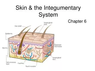
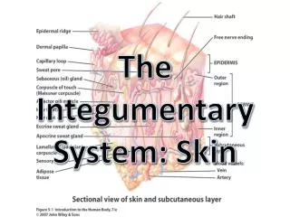
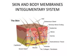
Two main layers and one below* • Epidermis - Outer layer - Composed of stratified squamous epithelium • Dermis • Inner layer • Thicker than epidermis • Made up of connective tissue, smooth muscle tissue, nervous tissue, blood, and other types of epithelial tissue (glands for example) *Beneath the dermis is the subcutaneous layer The epidermis and dermis are separated by the basement membrane that the stratified squamous epithelium is attached to
EPIDERMIS • Lacks blood vessels • Made up of 4 or 5 layers • Stratum Basale (closest to basement membrane) • Stratum Spinosum • Stratum Granulosum • Stratum Lucidum (Optional) • Stratum Corneum As each layer gets pushed upward, the cells change their shape and become more squamous or flattened.
Stratum Basale • The deepest layer – the contains cells that are actively dividing • Attached to the basement membrane • They are closest to the dermis, which contains blood vessels, so they receive nutrients and O2 • As they divide, they push the older cells toward the top. Cell division prevents wear and tear – the more the use of the body part, the more the cell division (calluses, corns, etc.) • Specialized cells in this layer called melanocytes, produce melanin, which provides skin color
Stratum Spinosum Stratum Spinosum • A thick layer of squamous epithelial cells right above the stratum basale
Stratum Granulosum Stratum Granulosum • A granular layer of squamous epithelium above the stratum spinosum
Stratum Lucidum Stratum Lucidum • An optional layer of squamous epithelium – only found in thick skin • Found in palms of hand or soles of feet
Stratum Corneum • Topmost layer of squamous epithelium. Flatten cells • These cells are called Keratinocytes, because their cell membranes thicken with a protein called keratin. • This hardens them, makes them waterproof. • The farther the keratinocytes travel from the stratum basale, the less nutrients they have, so they eventually die. • The older keratinocytes develop many desmosomes • The tough sheet of dead cells is called the Stratum Corneum and is shed (exfoliation)
Melanocytes Layers of the Epidermis
Melanocytes and Melanin • Although melanocytes are found in the stratum basale, their product – melanin can be found in other cells of the epidermis and sometimes even in the cells of the connective tissue underneath • This is because the melanocytes transfer the melanin to the neighboring cells via cellular extensions called dendrites – this process is called cytocrine secretion
More about Melanin • Melanin absorbs UV radiation – prevents skin cancer • Skin color depends mainly on the amount of the brown pigment melanin produced in the skin. • All people have about the same number of melanocytes. However, the melanocytes of dark-skinned people produce more melanin than do those of light-skinned people. The amount of melanin produced in each person's skin is determined mainly by heredity. • Exposure to sunlight increases the production of melanin, causing light skin to tan. In some cases, melanin builds up in small spots, forming freckles. • As someone grows older, the melanocytes produce melanin at uneven rates, which causes some areas of the skin to remain light and others to darken. These dark spots are sometimes called age spots or liver spots.
Sunburn (Solar Erythema) • Too much UV radiation • Epidermal cells become “sick” • Skin becomes red, swollen and painful • Cell undergo Apoptosis or programmed cell death • Apoptosis kills damaged cells to prevent skin cancer • Stratum basale will replace lost cells
Skin Cancer • Over exposure to UV light and other genetic and environmental conditions will cause skin cancer • Thymine dimers (T-T) form in the DNA of DNA repair enzyme genes • So DNA damage, misspellings cannot be proof-read or repaired • Mutations develop, leading to cancer • This occurs in a rare genetic disorder called Xeroderma pigmentosum
The Dermis • Below epidermis • Boundary between epidermis and dermis is uneven due to dermal papillae that extend into ridges in the epidermis • This creates ridges and fingerprints on the palm of the hand and fingers • Fingerprints are unique and genetically determined
Dermis, cont’d. • Two types of nerve receptors in the dermis: • Pacinian corpuscles: stimulated by heavy touch or pressure • Meissner’s corpuscles: stimulated by light touch • Made up of irregular dense connective tissue containing collagen and elastic fibers • Contains smooth muscle, blood vessels, nerve tissue, hair follicles, sweat glands, and sebaceous glands.
Subcutaneous Layer • Below dermis • Composed of loose connective tissue • No clear boundary between dermis and hypodermis • Contains adipose tissue layer for insulation • Contains major blood vessels that form a network called the Rete cutaneum – these send smaller extensions into the dermis
Accessory Organs of the Skin • Hair follicles • Nails • Skin glands • Sebaceous glands • Sweat glands (a.k.a sudoriferous glands) • Eccrine • Apocrine
Hair Follicles • Follicles are present on all skin excepts palms, soles, lips, nipples • In some areas that grow hair, the hair may be fine, while in others it may be thick • A hair grows out of a deep tube that extends from the surface of the skin, all the way into the dermal layer – this is a follicle • Each hair follicle is also connected to sebaceous glands that produce oils and other secretions
Hair Follicle, cont’d. • The follicle contains the growing hair • The base of the hair is composed of actively dividing epidermal cells • These epidermal cells are nourished by dermal blood vessels that project into connective tissue that then project into the hair base – this vascularized connective tissue projection is called the hair papilla
Hair Growth • As the epidermal cells divide, they push older cells upward through the follicle • As the older cells move away from the nutrient supply, they become keratinized and die • These dead keratinized epidermal cells are what make up a strand of hair • Hair color is genetically determined and is pigmented by melanocytes - the more the melanin in the dead epidermal cells, the darker the hair
A strand of hair has three layers: cuticle, cortex, and medulla. The outermost covering, cuticle consists of hard overlapping protein scales that point toward tips end. The cortex contains pigment granules that give hair its color. The medulla is a hollow tube within the hair, which may be present or absent. Depending on the hair, this hollow tube is continuous, or fragmented.
Hair Color • White – lacks melanin (albinism) • Gray hair – a mix of pigmented and unpigmented hair • Black and brown – tons of melanin • Blonds – Less melanin • Red hair – contains an extra pigment called trichosiderin (iron based)
Goosebumps! • Smooth muscle attached to each hair follicle can contract and cause short hairs to stand on end • These muscle bundles are called arrector pili muscles • They appear when scared, excited, cold, etc.
Nails • Evolved to protect the tips of fingers and toes • Consists of: • Nail plate (the actual nail) • Nail bed (skin surface on which the nail plate sits) • Nail plates are synthesized by special epithelial cells at the base of the nail – a region called a Lunula (half moon shaped) • As epithelial cells divide, they become keratinized, hard and scaly, and get pushed over the nail bed
Good Nails, Bad Nails Psoriasis of the nails Nail Biting
Glands - Sebaceous • Holocrine glands • Usually associated with hair follicles • Specialized epithelial cells in this gland produce a fatty secretion that accumulates and bursts through cells • The final product is a mix of cell debris and fatty fluid – this is called Sebum • Sebum is secreted into hair follicles • Keeps skin and hair soft, flexible and waterproof • Too much sebum causes pores to clog and results in acne
Glands - Sweat • Eccrine sweat glands • Most numerous on body • Eccrine ducts are independent of hair follicles • Respond to heat – secrete sweat to cool body • Fluid is mostly water, salt, urea and uric acid • Reaches surface of skin through a pore
Glands - Sweat • Apocrine sweat glands • Only found in armpits, groin, nipples • Apocrine ducts open into hair follicles • Become active during puberty • Secrete when excited, upset, anxious, etc. • Apocrine sweat is also basically water and salts, but also contains sebum and other fats that bacteria love and metabolize – this creates the bad odor in arm pits, etc.
Temperature Regulation • Humans (Mammals) are homeotherms ( warm-blooded). This means we maintain a fairly constant body temperature • We do this mainly by adjusting our rate of cellular respiration and by using organs such as the skin to warm and cool us down
Heat loss is caused by: • Radiation – heat from body radiates to cooler surroundings • Conduction – heat from body transfers directly to a cooler surface (your rear end warms your seat, so it feels warm to the touch) • Convection – heat from the body escapes to cooler air around the body • Evaporation – when sweat evaporates, it takes body heat away with it, cooling the body
Heat Loss, cont’d. In cold weather, blood vessels constrict, so that heat is retained in the body. In hot weather, blood vessels dilate, so heat is lost by convection and radiation Evaporative cooling As water molecules evaporate, they carry some of the heat with them, thus cooling the surface they leave behind
Hyperthermia • Normal body temp – 37C or 98.6F • Body temperature can get elevated during hot days and physical exercise • Body also elevates temperature (fever) to fight infection, because some bacterial and viral enzymes are denatured at above normal temperatures • But extremely high fevers can cause: • Dehydration and loss of electrolytes • the body’s own enzymes and proteins to denature and lead to death
Cool it! • Body temperature is reduced by sweating, staying in a cool environment and slowing physical activity • Drinking plenty of fluids • Fevers can be reduced by taking ibuprofen and acetaminophen
Hypothermia • Caused by over exposure to cold temperatures • Starts with shivering, followed by mental confusion, numbness, lethargy, loss of consciousness and eventually death
Accidents • Cuts • Burns
Clots When an injury such as a cut occurs, platelets become activated platelets. They change their shape, become sticky and build up on a blood vessel wall to form a plug. Platelets are also involved in the secretion of chemical platelet factors into the blood plasma. In a complex series of reactions fibrinogen is converted into fibrin, an insoluble protein that forms an intricate network of tiny threads called fibrils. Blood cells and plasma are tangled in the network of fibrils to form the clot.