Download
1 / 21
210 likes | 335 Vues
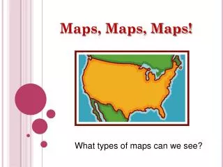
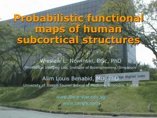
Functional Diffusion Maps (fDMs) for Brain Tumor Treatment Response Monitoring. Benjamin M. Ellingson, Ph.D. Assistant Professor of Radiology Dept. of Radiological Sciences David Geffen School of Medicine at UCLA.

E N D
Functional Diffusion Maps (fDMs) for Brain Tumor Treatment Response Monitoring Benjamin M. Ellingson, Ph.D. Assistant Professor of Radiology Dept. of Radiological Sciences David Geffen School of Medicine at UCLA B.M. Ellingson, Ph.D., Dept. of Radiological Sciences, David Geffen School of Medicine at UCLA, 2011
Diffusion MRI is sensitive to tumor cell density • Clinical ADC and cell density are negatively correlated • (Sugahara, 1999; Lyng, 2000; Chenevert, 2000; Gaurain, 2001; Nonomura, 2001; Guo, 2002; Chen, 2005; Hayashida, 2006; Manenti, 2008; Kinoshita, 2008; Ellingson, 2010) • ADC (or mean diffusion) = cell density (“hypercellularity”) • ADC = cell density (“hypocellularity”) Edema Necrotic Core Viable Tumor (Dark) ADC Map B.M. Ellingson, Ph.D., Dept. of Radiological Sciences, David Geffen School of Medicine at UCLA, 2011
ADC correlates with brain tumor cell density • Untreated glioma patients (WHO II-IV) underwent diagnostic stereotactic biopsy • (n = 17) biopsy sites were spatially matched to the pre-operative ADC map R2 = 0.7933; P < 0.001 ADC Sensitivity = 1.01 x 10⁻⁷ [mm2/s]/[nuclei/mm2] B.M. Ellingson, Ph.D., Dept. of Radiological Sciences, David Geffen School of Medicine at UCLA, 2011 From: Ellingson BM et al., J Magn Reson Imaging, 2010
The Functional Diffusion Map (fDM)(Moffat, 2005; 2006; Hamstra, 2005; 2008; Ellingson, 2009; 2010; 2011) From: Ellingson, JMRI, 2009, In Press B.M. Ellingson, Ph.D., Dept. of Radiological Sciences, David Geffen School of Medicine at UCLA, 2011
The Functional Diffusion Map (fDM)(Moffat, 2005; 2006; Hamstra, 2005; 2008; Ellingson, 2009; 2010; 2011) From: Ellingson, JMRI, 2009, In Press B.M. Ellingson, Ph.D., Dept. of Radiological Sciences, David Geffen School of Medicine at UCLA, 2011
Early Detection of Brain Tumor Growth T1+C Contrast-Enhancement (white) FLAIR fDMs B.M. Ellingson, Ph.D., Dept. of Radiological Sciences, David Geffen School of Medicine at UCLA, 2011
fDMs in Brain Tumor Progression 3 mo. 6 mo. 9 mo. (Onset of symptoms) T1+C FLAIR fDM B.M. Ellingson, Ph.D., Dept. of Radiological Sciences, David Geffen School of Medicine at UCLA, 2011
fDMs in Progressive Disease (PD) Hypercellularity Hypercellularity Hypercellularity From: Ellingson, ISMRM, 2009; SNO, 2009 B.M. Ellingson, Ph.D., Dept. of Radiological Sciences, David Geffen School of Medicine at UCLA, 2011
fDM Results in Stable/Responding Disease (SD/RD) Hypocellularity Hypocellularity Hypocellularity From: Ellingson, ISMRM, 2009; SNO, 2009 B.M. Ellingson, Ph.D., Dept. of Radiological Sciences, David Geffen School of Medicine at UCLA, 2011
fDMs May Reflect Molecular/Genetic Phenotypes MGMT(+) MGMT(-) MGMT(+) MGMT(-) MGMT(+) MGMT(-) From: Ellingson, ISMRM, 2009; SNO, 2009 B.M. Ellingson, Ph.D., Dept. of Radiological Sciences, David Geffen School of Medicine at UCLA, 2011
fDMs May Reflect Molecular/Genetic Phenotypes From: Ellingson, ISMRM, 2009; SNO, 2009 B.M. Ellingson, Ph.D., Dept. of Radiological Sciences, David Geffen School of Medicine at UCLA, 2011
Clinical fDM Sensitivity/Specificity WHO Grade (n = 50 Total Patients) Spearman Corr. Coeff. R = 0.4350, P = 0.0016 B.M. Ellingson, Ph.D., Dept. of Radiological Sciences, David Geffen School of Medicine at UCLA, 2011 From: Ellingson BM et al., ISMRM, 2010
Clinical fDM Sensitivity/Specificity Neurological Status (n = 50 Total Patients) Pearson Corr. Coeff. R2 = 0.8586, P < 0.0001 B.M. Ellingson, Ph.D., Dept. of Radiological Sciences, David Geffen School of Medicine at UCLA, 2011 From: Ellingson BM et al., ISMRM, 2010
fDMs Before/After Radiotherapy N = 94 B.M. Ellingson, Ph.D., Dept. of Radiological Sciences, David Geffen School of Medicine at UCLA, 2011
fDMs Before/After Bevacizumab N = 77 B.M. Ellingson, Ph.D., Dept. of Radiological Sciences, David Geffen School of Medicine at UCLA, 2011
Graded fDMs B.M. Ellingson, Ph.D., Dept. of Radiological Sciences, David Geffen School of Medicine at UCLA, 2011
Graded fDMs:Tumor Invasion Beyond FLAIR B.M. Ellingson, Ph.D., Dept. of Radiological Sciences, David Geffen School of Medicine at UCLA, 2011
Graded fDMs for StealthStation™ Biopsy Localization B.M. Ellingson, Ph.D., Dept. of Radiological Sciences, David Geffen School of Medicine at UCLA, 2011
Graded fDMs in Differential DiagnosisTumor vs. Demyelination Hypercellular Hypocellular Macrophages & Inflammatory Cells Demyelination Biopsy Diagnosis = Demyelination (Multiple Sclerosis) B.M. Ellingson, Ph.D., Dept. of Radiological Sciences, David Geffen School of Medicine at UCLA, 2011
Graded fDMs – Radiation Necrosis T1+C FLAIR Hypercellular Graded fDM Hypocellular B.M. Ellingson, Ph.D., Dept. of Radiological Sciences, David Geffen School of Medicine at UCLA, 2011
Stereotactic fDMs Using a Neuroanatomical Atlas Overlaid On T1 Atlas Anatomy Label Map B.M. Ellingson, Ph.D., Dept. of Radiological Sciences, David Geffen School of Medicine at UCLA, 2011