Download
1 / 19
200 likes | 421 Vues
Overview of 2DE. Complex mixture of proteins. Denature and solubilize in solution (Sample prep). Separate proteins by charge in first dimension (IEF). Separate proteins by size in second dimension (SDS-PAGE). Individual proteins isolated as distinct protein features within a gel matrix.

E N D
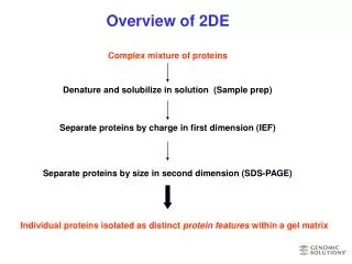
Overview of 2DE Complex mixture of proteins Denature and solubilize in solution (Sample prep) Separate proteins by charge in first dimension (IEF) Separate proteins by size in second dimension (SDS-PAGE) Individual proteins isolated as distinct protein features within a gel matrix
Non-ionic and zwitterionic detergents Sample Buffer I Remove non-protein components Centrifugation and degrading enzymes Sample Preparation Procedure GSI products Disrupt tissues and cell membrane Mechanical force and detergents Sample Buffer II RNAse and DNAse Disrupt complexes and ‘linearize’ proteins chaotropic agents (Urea) reducing agents (DTT) detergents Rehydration buffer IPG loading buffer Sample buffer III Eliminate highly abundant proteins Cibachron dye mini-columns Remove salt from fractionated or affinity purified samples Dialysis kits and buffers
Isoelectric Focusing- Separation by Charge Anode Cathode - + pH 3 4 5 6 7 8 9 10 Acidic Basic Proteins are amphoteric (contain acidic and basic residues) For every protein there is a pH at which its net charge is 0. This is its isoelectric point (pI) Above its pI, a protein has an overall negative charge and will migrate toward the positively charged anode Below its pI, a protein has an overall positive charge and will migrate toward the negatively charged cathode At its pI, a protein does not move (focuses into a single band)
Final Result of IEF BPB Acidic Basic Proteins focused into distinct bands
Methods for isoelectric focusing Carrier-ampholyte tube gels pH gradient created by discontinuous buffering system and carrier ampholytes Immobilized pH Gradient(IPG) strips pH gradient fixed in gel by covalently linking amphoytes to acrylamide when gel gradient is poured.
Investigator Tube Gel Apparatus Capacity: 15 analytical or 8 preparative tubes
Investigator IPG pHaser Investigator IPG pHaser Runs up to 10 IPG strips Compatible with all brands of IPG strips
Tube gels vs IPG strips Benefits of tube gels No rehydration step- saves one day. May be better for some proteins-membrane, hydrophobic. Benefits of IPG strips Immobilized pH gradient eliminates cathodic and anodic drift. Higher volume of sample can be loaded. Less likely to become damaged in 2DE procedure. Less labor involved.
Second dimension SDS Page- Separation of proteins by mass Coat focused proteins with SDS GSI products: Equilibration buffers I and II Place strip/tube directly onto 2nd D gel GSI products: 2-D Running System-tank, power supply, chiller, precast slab gels, gel casting reagents, premixed buffers Negatively charged proteins migrate toward + anode Anode +
Typical results following 2DE High MW Low MW Acidic Proteins Basic Proteins
Investigator 2DE Electrophoresis System Peltier chiller Programmable Power Suppy Capacity: 5 single or 10 double gels Single power supply runs tube gels, IPG strips and slab gels
Detection of protein features Detection limits Staining method Standard Coomassie Colloidal Coomassie Fluorescent Dyes Non-destructive Silver Destructive Silver 300ng 30ng 5ng 5ng 1ng
Standard Coomassie Staining General method Add stain with fixative and incubate 4hrs to overnight. Destain in 40% Methanol, 10% acetic acid. Benefits Easy and consistent Mass-spec friendly Disadvantages Least sensitive stain Requires long incubation times and destaining
Colloidal Coomassie Staining General Method Incubate gel in colloidal solution for minutes or hours Destain with water (if required). Benefits Mass spec friendly Very fast Environmentally friendly/ less hazardous More sensitive than standard coomassie No special visualization requirements Disadvantages Detection limit 10-50 times lower than fluorescent dyes or silver.
Staining using Fluorescent dyes General method Incubate in dye for 1 hour to overnight Destain (if desired) Benefits Fast and easy to use Non-toxic Very sensitive Linear over a broad range (ng to mg) Mass spec friendly Disadvantages Requires UV source for visualization
Pros and Cons of Silver Staining Benefits Most sensitive stain (when gluteraldehyde is used) Disadvantages Long and tedious procedure Labor intensive Hazardous Very sensitive to wash and development times Linear over a very narrow range Protein specific staining (some do not stain or stain negatively) Most sensitive method (destructive) not compatible with mass spec
How much sample should I load? The amount of sample required depends on both the staining method, AND the complexity of the sample Examples: If the sensitivity for Coomassie equals 40-50ng per feature, then 1,000 features could be detected starting with 50-100ugs sample. To visualize all the features in a sample containing 10,000 features you need to start with 500ug-1mg total protein. If the sensitivity for silver equals 1-5ng per feature, then 1000 features could be detected starting with 1-5ug sample. To visualize all the features in a sample containing 10000 features you need to start with 10-50ug total protein. Note: These examples assume that all proteins in the sample are present in equal amounts, not the case in real life.
Finding what you’re looking for The ‘simple’ eukaryote, yeast, has approximately 6000 genes but can produce over 12,000 protein features due to post-transcriptional and post translational modifications. To ‘see’ a rare protein within a crude extract, 20-2000mgs of total protein would need to be loaded onto the 1st dimension gel. Therefore 2DE of crude fractions using broad range IEF gels can only provide information on relatively abundant proteins (high copy number). If you want to detect moderate to rare proteins you must reduce the complexity of the sample or limit the scope of the search.
2-D gel of sample before (left) and after (right) treatment with albumin depletion kit. Methods for finding the ‘interesting’ proteins Prefractionate- Reduce the complexity of each sample by fractionating into nuclear, cytoplasmic, mitochondial, microsomal or other distinct compartments. Remove highly abundant proteins Purify complexes- Enrich for specific activities, or complex components. Use narrow range IEF gels- Increase the amount of sample you can load on a gel while increasing resolution within narrow PI ranges (3-6, 5-7,6-8,7-9). The use of zoom gels (one point pH spread) allows loading of up to 40mgs starting sample.